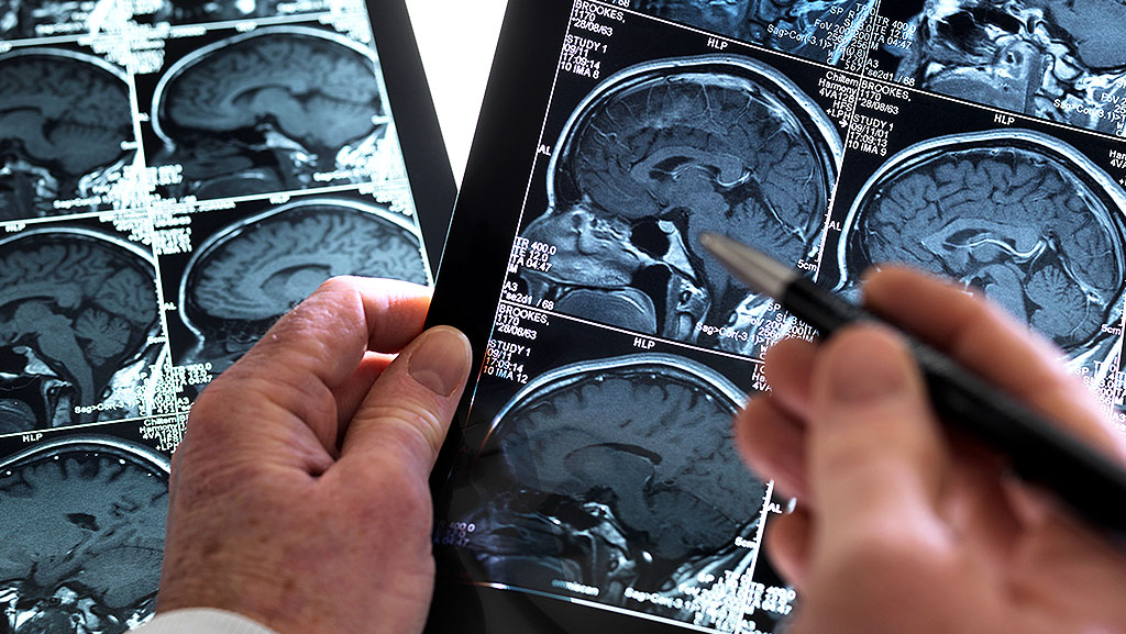
MRI and fMRI are two types of magnetic resonance imaging that doctors and scientists use to look inside the body and the brain in great detail. Both scans use a powerful MRI scanner magnet to produce images without harmful X-rays or ionizing radiation, making them safe and noninvasive ways to see inside the human body. The MRI provides a clear picture of the body’s anatomical structure, such as tissues and organs, while the fMRI shows how parts of the brain function by tracking blood flow and activity over time.
People often feel confused when they hear the terms “MRI” and “fMRI,” because the names are similar. This article explains the key differences between these two scans in very simple English, so you understand what eachdoes, how each works, and why one might be used instead of the other. The goal is to make it easy for everyone to learn the difference between MRI and fMRI.
What Is MRI? (Magnetic Resonance Imaging)
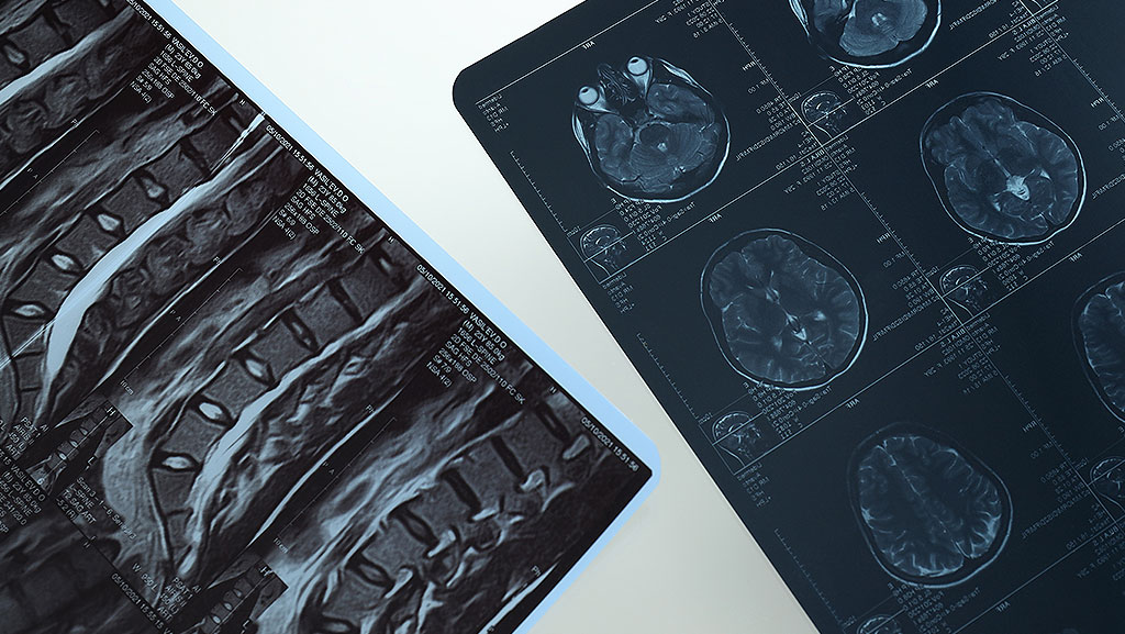
Magnetic Resonance Imaging, or MRI, is a type of scan that creates very detailed, high-resolution images of the inside of your body. It shows soft-tissue contrast of organs, muscles, and other structures in areas such as the brain, spine, heart, and joints. MRI uses a strong magnetic field and radio waves to make pictures of the body. It does not use radiation, so it is safer than X-rays or CT scans for repeated imaging.
When you get an MRI scan, you lie still inside a big machine that has a large magnet around you. The machine aligns the tiny hydrogen atoms in your body and uses radiofrequency coils to collect signals. A computer then turns these signals into clear images of your body’s internal structures. You do not feel the magnet, but the machine can be loud, and you must stay still for the best results.
Doctors use MRI scans to look for many things. MRI is helpful for detecting conditions such as tumors, injuries, and conditions affecting the brain, spinal cord, and other organs. Because it provides detailed pictures of organs and soft tissue, it helps doctors diagnose disease, plan treatments, and monitor changes over time.
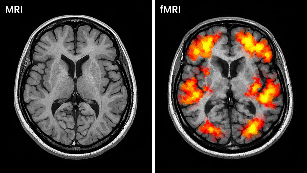
MRI focuses on structural MRI and shows what the body looks like right now. It does not show how parts of the brain are working or what they are doing at the moment. That is where fMRI becomes useful.
What is fMRI (Functional MRI)
Functional MRI, or fMRI, is a specialized MRI scan that shows how the brain works by tracking blood flow in real time. It uses the same strong MRI scanner magnet and radiofrequency coils as a normal magnetic resonance imaging test, but the key difference is that it does more than show structure. fMRI maps the active parts of the brain when you do tasks or think about something.
When parts of your brain become active, they use more oxygen. This leads to local changes in blood oxygenation that fMRI can detect. The scan records these signals and creates moving functional imaging maps that show brain activity and how different brain regions work together. This is known as the Blood Oxygen Level Dependent or BOLD signal.
Doctors and scientists use fMRI for functional brain mapping and for neuroscience research. It helps with neurosurgery planning, studying memory and language, and understanding conditions like epilepsy or stroke by showing which parts of the brain control certain actions or functions.
Unlike anatomical MRI, which produces static images of organs and tissues, fMRI produces dynamic images of brain activity. This is useful in both research studies and in some clinical settings, where understanding how the cerebral cortex or other brain regions work is important.
MRI vs fMRI — Side-by-Side Comparison
Let’s see the key differences between MRI and fMRI in simple English so you can easily see what each scan measures and why doctors choose one over the other.
What They Measure
MRI produces static images of the body’s shape and anatomical structures, such as organs, tissues, and soft tissues, with soft-tissue contrast. It shows a “snapshot” of what the body looks like at one point in time. fMRI makes dynamic maps of brain activity by tracking changes in blood flow and oxygen as brain regions work. This means MRI focuses on structure, while fMRI focuses on function.
Type of Information Provided
- MRI provides detailed images used to diagnose structural problems such as tumors, injuries, or organ abnormalities.
- fMRI produces moving images that show which brain areas are active during tasks or at rest, using the Blood Oxygen Level-Dependent (BOLD) signal.
Spatial vs Temporal Resolution
MRI has high spatial resolution, allowing it to show small details clearly. fMRI has higher temporal resolution, meaning it tracks changes in brain activity over time. This lets fMRI show how brain activity shifts from moment to moment.
Patient Experience and Tasks
During an MRI scan, you simply lie still inside the machine while it makes detailed images of your body. During an fMRI scan, you may be asked to perform simple tasks, such as thinking, tapping your hand, or looking at pictures, so the scan can measure brain activity in response to those actions.
Uses in Medicine and Research
- MRI is widely used for diagnostic imaging to detect structural abnormalities such as injuries, strokes, or diseases in many parts of the body.
- fMRI is often used in brain research and in planning neurosurgery to map active brain areas, such as those responsible for speech, movement, or thinking.
Availability and Cost
MRI machines are common in many hospitals and clinics. fMRI scans are less common, more expensive, and typically conducted in research hospitals or centers with specialized equipment and software.
Summary of Key Differences
| Feature | MRI | fMRI |
| What it shows | Detailed structure | Active brain function |
| Image Type | Still images | Dynamic maps |
| Used for | Diagnosing physical issues | Brain activity research and planning |
| Tasks during scan | Stay still | May perform simple tasks |
| Common use | Widely available | Less common and more costly |
Knowing these differences helps you understand what each scan does and why a doctor might order one instead of the other. MRI provides a clear picture of the body, while fMRI shows how the brain works.
Why Understanding the Difference Matters
Understanding the difference between MRI and fMRI helps you know when and why each scan is used. MRI and fMRI both use strong magnetic fields and radio waves to make images of the body, but they provide very different information. MRI provides detailed structural images, while fMRI shows brain activity and function over time. Knowing this helps you understand medical advice and what the scan can do for you or a loved one.
Doctors choose MRI vs fMRI based on what they need to learn. If a doctor needs to visualize anatomical structures such as organs, joints, or tissues, they use an MRI. This helps detect problems such as tumors, injuries, or changes in organ shape.
If doctors or researchers want to know how the cerebral cortex and other brain regions work during thinking, movement, or memory tasks, they use fMRI. fMRI maps blood flow and uses the Blood Oxygen Level Dependent signal to show which parts of the brain are active during those tasks.
For patients, knowing the difference also sets clear expectations for the scan experience and results. Patients learn why they may be asked to do simple tasks during an fMRI and why an MRI might be shorter or just involve lying still.
In research and clinical settings, understanding both scans helps combine structural MRI and functional imaging data to get a more complete picture of a patient’s health. This can lead to better diagnosis, more effective treatment planning, and deeper insights into conditions such as stroke and neurological disorders.
Practical Scenarios: When Each Scan Is Used
In this section, we explain real-world uses of MRI and fMRI in clear, simple language so you understand when and why doctors and researchers choose each scan. Both scans are types of magnetic resonance imaging, but they serve different purposes in medical imaging and in neuroscience research.
Classic Uses for MRI
MRI scans are powerful tools for evaluating the structure of the body. They are widely used in hospitals and clinics because they show detailed anatomy of organs, bones, and tissues without radiation. Doctors use MRI for many tasks:
- Brain structure checks for conditions like tumors, strokes, or tissue damage.
- Spinal issues such as disc problems or nerve compression.
- Joint and muscle injuries may show tears or inflammation.
- Heart and blood vessel imaging to assess heart structure.
MRI delivers high spatial resolution images, meaning it shows fine detail in the body’s structure. Doctors often choose MRI when they want a clear snapshot of the body’s current appearance.
When fMRI Is Used
fMRI scans are mainly used to see how the brain works rather than just its shape. fMRI measures dynamic changes in blood flow and brain activity during thinking, movement, or task performance. This makes fMRI valuable in scenarios such as:
- Neurosurgery planning to map important brain areas, such as speech or movement regions, before an operation.
- Stroke recovery studies show how parts of the brain reorganize after injury.
- Brain research on memory, language, emotion, and learning.
- Psychiatric and neurological research to explore functional connectivity and brain networks.
Unlike MRI, fMRI captures brain function over time using the Blood Oxygenation Level Dependent (BOLD) signal to infer which brain regions are active during tasks.
Combined Use in Medicine and Research
In some clinical settings, doctors use both MRI and fMRI together. For example:
- Before complex brain surgery, an MRI gives the detailed structure of the brain and fMRI maps the active functions near risky areas.
- Neurodegenerative disease studies may use MRI to see degeneration and fMRI to study changes in brain activity.
This combined use helps provide a complete picture of brain health and guides treatment planning more accurately than either scan alone.
Summary of Practical Uses
- MRI: Best for diagnosing structural conditions, like tumors, injuries, or physical abnormalities.
- fMRI: Best for understanding brain function, such as activity during thinking, behavior tasks, or planning surgery.
These examples show how MRI and fMRI are used in everyday medicine and research to answer different questions about the body and the brain.
Safety & Patient Experience
MRI and fMRI are both types of magnetic resonance imaging that use very strong magnetic fields and radiofrequency coils to produce images of the body or brain without harmful radiation. This makes both scans safer than X-rays or CT scans, which use ionizing radiation. MRI is generally safe, but patients must follow specific guidelines to avoid risks associated with metal and other conditions.
Both scans require you to lie very still inside a large tube-like machine while it makes images. The machine is quiet but can make loud banging noises, so you will often wear ear protection. Remaining still is very important because even small movements can blur the images.
Before either scan, you will fill out a screening form to make sure you do not have any metal objects or electronic devices in or on your body, such as pacemakers, implants, or shrapnel. These items could move or malfunction due to the strong magnet, posing a risk of injury. Jewelry, watches, or metal clothing parts should be left at home or removed before the scan.
Some patients feel claustrophobic or uncomfortable inside the scanner’s tunnel, so doctors may give medication to help you relax. Pregnancy does not automatically prevent you from having an MRI or fMRI, but doctors will decide whether the scan is necessary, especially in early pregnancy.
During an fMRI scan, you may be asked to do simple tasks like tapping your fingers, looking at pictures, reading, or listening to sounds so the scan can map brain activity. This is different from a normal MRI, where you usually lie still without tasks.
Overall, both MRI and fMRI are noninvasive and safe for most people, but it is important to talk to your doctor and MRI technologist about any health conditions, implants, or concerns you have before the scan.
FAQs (Frequently Asked Questions)
Here are clear answers to common questions people ask about MRI and fMRI scans. This section uses simple English to help you understand what each scan does and what to expect.
What is an MRI?
MRI stands for magnetic resonance imaging. It is a safe, non-invasive scan that uses a powerful magnet and radio waves to make detailed images of the inside of your body, like organs, tissues, and the brain’s structure. It does not use harmful X-rays.
What is an fMRI?
fMRI stands for functional magnetic resonance imaging. It is a type of MRI scan that shows how parts of your brain work by measuring changes in blood flow as your brain becomes active. This helps create functional maps of brain activity.
How are MRI and fMRI different?
A regular MRI produces a still image of structures such as organs and tissues. fMRI shows how the brain is functioning over time by tracking changes in oxygen and blood flow.
Will I feel pain during the scan?
Most people do not feel pain during either MRI or fMRI. You may hear loud noises from the machine, so stay very still. If you have questions or concerns, be sure to speak with the technologist before your scan.
Do these scans use radiation?
No. MRI and fMRI do not use radiation like X-rays or CT scans. They use magnetic fields and radio waves instead, which are generally safe.
Will I need contrast dye?
Some MRI tests use contrast dye to make certain tissues easier to see. For fMRI, contrast is usually not required unless your doctor says it is needed.
Can anyone have an MRI or fMRI?
Most people can have these scans, but if you have any metal or electronic devices in your body (such as certain pacemakers), you must tell your doctor. These devices can be unsafe inside the strong magnetic field.
Why might a doctor choose fMRI instead of MRI?
A doctor may choose fMRI when they need to see how the brain functions during tasks such as movement or speech, or when planning neurosurgery. A regular MRI is chosen when the goal is to look at structure and anatomy.
Can fMRI show mental health conditions?
fMRI can help research how the brain works in conditions like memory problems or brain injury, but it cannot diagnose mental illness on its own.
How long does each scan take?
MRI and fMRI scans can take from about 15 minutes to more than an hour, depending on what the doctor needs to see. During fMRI, you may also be asked to do simple tasks so the scan can map your brain activity.
Conclusion
In simple terms, MRI and fMRI are two important types of magnetic resonance imaging that help doctors and researchers see inside the body and brain in very different ways. An MRI scan provides detailed images of internal structures, such as organs, tissues, and bones. It provides a clear snapshot of the body at a single moment, making it very useful for diagnosing structural problems such as injuries, tumors, and issues with the spine and joints. MRI does not use radiation, so it is safe and widely used in many medical settings.
On the other hand, fMRI is a type of MRI that measures brain activity by detecting changes in blood flow and oxygen. It creates dynamic maps that show which parts of the brain are working during tasks or when at rest. This makes fMRI useful for neuroscience research, neurosurgery planning, and understanding how the brain’s functional networks work. fMRI does not replace MRI for structure, but it adds another layer of information about function and activity.
Both scans are noninvasive and safe for most people, and each one plays a special role in medical imaging, clinical MRI applications, and brain research. Choosing between MRI vs fMRI depends on what question the doctor or researcher needs to answer: “What does the body look like?” or “How is the brain working?” Understanding the difference helps you know why one scan is used over the other and what to expect from the results.