Interventional pain procedures are medical treatments that help physicians identify and treat the source of pain within the body. These procedures employ image guidance to improve treatment safety and accuracy. Image guidance enables physicians to visualize the body’s anatomy, allowing them to place needles, catheters, or other instruments in the correct position and to avoid injury to adjacent structures. Without imaging, doctors might have to guess where to put the needle based only on feel or surface marks. Modern imaging makes this guesswork much safer and more exact.
Imaging guidance uses modalities such as fluoroscopy, ultrasound, CT, MRI, and X-ray to provide real-time images of the area being treated. These technologies help physicians target sites such as the epidural space, spinal nerve root, facet joint, sacroiliac joint, and peripheral sensory nerve during procedures, including epidural steroid injections, facet joint injections, peripheral nerve blocks, and radiofrequency ablation.
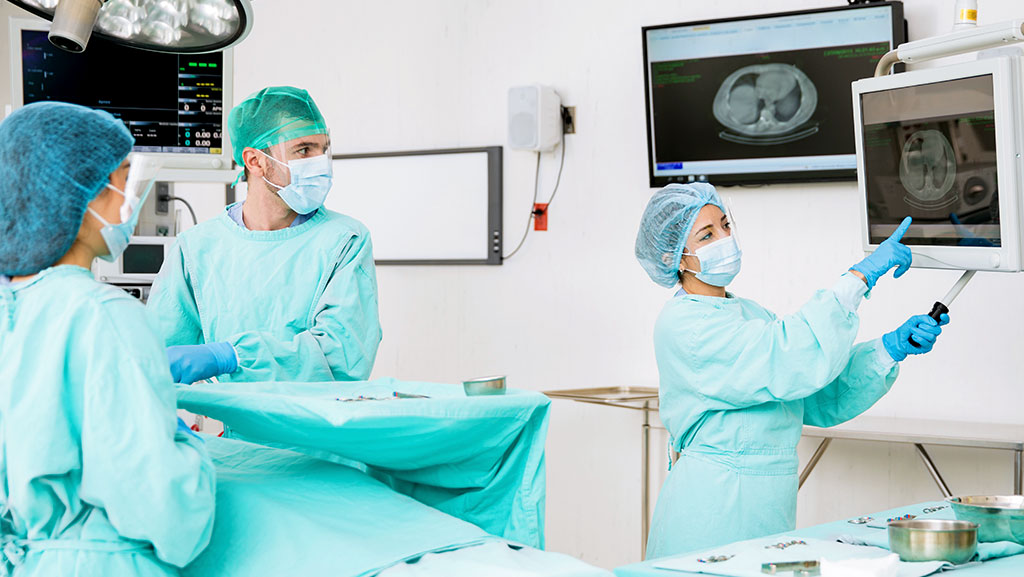
Doctors such as pain management specialists, interventional radiologists, ultrasound technicians, and radiologic technologists collaborate to use these guidance tools. Imaging guidance improves needle placement accuracy, reduces complications, and increases the chances of successful pain relief. Patients with chronic pain conditions such as chronic low back pain, sciatica, degenerative disc disease, and osteoarthritis may benefit from these image-guided interventions.
What Is Image-Guided Interventional Pain Management
Image-guided interventional pain management means physicians use imaging to perform pain procedures more accurately, safely, and with better outcomes than without imaging. These methods allow physicians to visualize the body while guiding a needle, a tool, or medication to the source of pain, rather than relying on touch or surface marks to infer location.
Most interventional pain procedures today use real-time imaging to show the anatomy before and during the treatment. This imaging helps place the needle in the correct portion of the epidural space, near a spinal nerve root, into a facet joint, or close to a peripheral sensory nerve with fewer errors.
Using imaging technologies such as fluoroscopy, ultrasound, and CT provides a clear picture of bones, joints, and soft tissues, enabling physicians to carefully guide injections of pain medication or blocks to the exact area requiring treatment. This makes procedures like epidural steroid injections, facet joint injections, and peripheral nerve block safer and more precise.
Doctors such as pain management specialists, interventional radiologists, and trained imaging staff have found that using imaging during these procedures reduces the risk of complications, helps confirm correct needle or instrument placement, and provides a clearer view of the anatomy when conditions are complex.
In contrast, procedures performed without imaging have higher rates of incorrect placement and a greater risk. Imaging guidance is now considered the standard for many spine and joint injections because it improves needle placement accuracy, reduces side effects, and helps doctors and patients have more confidence in the results.
Core Imaging Modalities and Their Roles
Doctors use various imaging modalities to guide interventional pain procedures, enabling visualization of the body and accurate targeting of the pain source. These tools help guide needles, medicines, and instruments in real time. Imaging makes procedures like epidural steroid injection, peripheral nerve block, and facet joint injection much safer than doing them without imaging. Most spinal and joint procedures now use imaging as the standard of care.
2.1 Fluoroscopy
Fluoroscopy is one of the most common imaging methods doctors use in pain management. It is a real-time X-ray guidance technique that helps physicians visualize bones and joint spaceswhile placing a needle or instrument. This is very helpful for spine injections and joint procedures because the spine’s bony structures are easy to see on fluoroscopy.
Fluoroscopy is often used for:
- Epidural steroid injections
- Facet joint injection
- Spinal nerve root targeting
Doctors may also use contrast dye with fluoroscopy to visualize the needle tip before medication administration.
2.2 Ultrasound
Ultrasound uses sound waves to show soft tissues, nerves, and blood vessels in real time. This imaging modality does not use ionizing radiation and helps physicians visualize nerve pathways and soft tissues that X-rays do not visualize well. As a result, ultrasound is increasingly used for nerve blocks and other pain procedures.
Ultrasound may be used for:
- Peripheral nerve block
- Soft tissue visualization
- Guiding needle placement near nerves or muscles
Ultrasound also allows physicians to avoid adjacent blood vessels, thereby reducing the risk of complications.
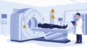
2.3 Computed Tomography (CT)
Computed Tomography (CT) produces detailed cross-sectional images of bones and tissue. CT can help doctors place needles in deep or complex body parts that are hard to see with other tools. It provides a clear image of the internal anatomy and is useful when precision is critical.
CT-guided injections are often used when physicians require greater detail than fluoroscopy or ultrasound alone can provide.
2.4 Magnetic Resonance Imaging (MRI)
MRI provides clear images of soft tissues, including nerves, discs, and muscles. It does not use radiation. In some pain procedures, physicians use MRI planning images to understand the anatomy before the procedure, and, in exceptional cases, MRI can also guide interventions.
MRI is helpful for:
- Deep soft tissue visualization
- Planning complex procedures
How Imaging Improves Precision and Safety
Imaging guidance significantly improves interventional pain procedures by enabling physicians to place needles and instruments more accurately and safely than when relying solely on tactile or body landmarks. When physicians can visualize the target area using fluoroscopy, ultrasound, CT, MRI, or X-ray guidance, they reduce the risk of needle placement in the wrong location. Studies show that without imaging, needle placement can be incorrect in many cases of spine injections, but imaging considerably lowers these risks.
Improved Accuracy
When doctors use imaging, they can see the exact spot where a needle should go. This helps deliver medications directly to the site of pain, such as near the spinal nerve root, the epidural space, or thefacet joint. Clear images from imaging tools enable physicians to aim precisely, so that injections such as epidural steroid injection or peripheral nerve block reach the correct target area the first time.
Better Safety
Seeing the needle and nearby anatomy in real time helps clinicians avoid injuring essential structures such as blood vessels or nerves. This lowers the chances of complications like bleeding or nerve damage, which can happen when doctors cannot see inside the body. Imaging also flags if a needle is accidentally placed in a blood vessel or disc space, so doctors can correct it before injecting medicine.
Reduced Complications
Using imaging reduces the need for repeat injections and improves procedural control. The real-time view of the procedure helps physicians guide needles into the correct position with fewer attempts. Fewer needle passes reduce the risk of infection and other complications, making procedures such as facet joint injection and nerve blocks safer.
Targeted Treatment
When imaging shows the exact location of the pain source, medicines can be delivered right where they are needed. This precise delivery helps the medication work more effectively. It can lead to faster and more lasting pain relief for conditions such as chronic low back pain, sciatica, degenerative disc disease, and osteoarthritis.
Patient Comfort and Confidence
Patients often feel more comfortable knowing the doctor can see inside their body during the procedure. Because imaging helps guide smaller needles and requires fewer needle passes, patients usually experience less pain during and after the procedure.
Common Image-Guided Pain Procedures
Image-guided pain procedures use modalities such as fluoroscopy, ultrasound, CT, and MRI to help physicians place needles, blocks, and medication precisely where the pain originates. Most common procedures are done under imaging guidance because it is considered the standard of care and improves safety and outcomes.
4.1 Epidural Steroid Injection
Epidural steroid injection is a procedure where doctors place medicine into the epidural space to calm irritated or inflamed spinal nerve roots. This can help reduce pain from conditions like sciatica or degenerative disc disease and may allow patients to move more easily after treatment. Imaging, such as fluoroscopy or CT, helps guide the needle so the medicine goes precisely to the right place.
4.2 Facet Joint Injection
Facet joint injection targets the small joints in the spine that help with movement. Pain in these joints can cause chronic low back pain or neck pain. A physician uses imaging, such as fluoroscopy or CT, to guide the needle into the correct facet joint before administering medication to reduce inflammation. This helps confirm that the joint is causing pain and provides targeted relief.
4.3 Peripheral Nerve Block
A peripheral nerve block is used to deliver medication near a group of nerves outside the spinal cord. These blocks can reduce pain in areas such as the arms, legs, or joints. Ultrasound guidance is commonly used for peripheral nerve blocks because it can visualize soft-tissue structures and nerves in real time. This helps the doctor avoid nearby blood vessels and improves accuracy.
4.4 Selective Nerve Root Block
A selective nerve root block targets a specific spinal nerve root that may be causing radiating pain. Imaging guidance, such as fluoroscopy, helps ensure that the needle reaches the correct nerve root before medication is injected. This procedure can also help confirm the source of pain for diagnostic purposes and for relief.
4.5 Sacroiliac Joint Injection
Sacroiliac joint injection treats pain that comes from the joint between the spine and pelvis. As with other procedures, imaging guidance helps place the needle accurately within the joint, enabling delivery of anti-inflammatory medication where it is needed.
4.6 Other Image-Guided Blocks and Injections
There are many other injections and blocks guided by imaging in interventional pain care, including:
- Trigger point injections targeting sore muscle spots
- Celiac plexus block for abdominal pain relief
- Radiofrequency ablation where imaging confirms placement of a heat-delivering probe near nerves
These image-guided procedures help treat pain with precise targeting and reduced risk of complications.
Choosing Between Imaging Modalities
When physicians decide how to guide an interventional pain procedure, they select the most appropriate imaging modality based on the patient’s anatomy, the procedure type, and the risks and benefits of each modality. No single imaging modality is optimal for every case, and each has its own role in guiding pain procedures safely and accurately.
Fluoroscopy vs. Ultrasound
Fluoroscopy is a mainstay imaging method in many pain clinics. It uses real-time X-ray guidance to show bones and joint spaces as the needle or instrument moves. Doctors often choose fluoroscopy for spine procedures like epidural steroid injection and facet joint injection because it clearly shows the bony anatomy and offers continuous feedback while the needle is placed.
Ultrasound does not use ionizing radiation and can visualize soft tissues, nerves, and blood vessels in real time. It allows physicians to observe the needle tip’s movement and see how medications spread around nerves or muscles. This is particularly useful for peripheral nerve blocks and soft-tissue injections. Ultrasound also reduces radiation exposure, so it may be a better choice for some patients.
In some cases, clinical studies have shown that ultrasound- and fluoroscopy-guided injections yield similar pain relief and safety outcomes. However, fluoroscopy remains preferred for some spine injections, whereas ultrasound is increasingly used for nerve blocks and joint injections, where soft-tissue detail is essential.
When CT Might Be Best
Computed Tomography (CT) offers very detailed cross-sectional images of bones and tissues. This can improve needle placement accuracy in complex or deep areas that are difficult to visualize with fluoroscopy or ultrasound alone. CT is often used when the joint or anatomy is irregular or complex to access, or when it is located deep within the body.
Although CT gives excellent detail, it may not be available in all pain clinics and usually exposes patients to more radiation than fluoroscopy. Thus, physicians reserve CT for cases where additional detail is necessary to guide the procedure safely.
Role of MRI in Guidance
MRI provides the best soft-tissue contrast among standard imaging modalities and does not use ionizing radiation. In interventional pain care, MRI planning images can help physicians understand complex anatomy before a procedure. It is not used as frequently during needle placement because MRI machines are larger and less accessible than fluoroscopy or ultrasound equipment.
Patient Benefits and Outcomes
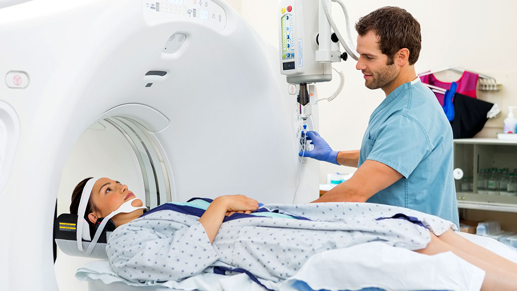
Image-guided pain procedures help many patients get better results than traditional injections without imaging. When physicians use imaging modalities such as fluoroscopy, ultrasound, CT, and occasionally MRI, the procedure becomes more precise, safer, and easier for patients to tolerate. Research shows that imaging can help reduce pain, improve function, and lower the chance of complications.
More Accurate Pain Relief
Using image guidance allows physicians to place medication precisely at the site of pain. This makes treatments such as epidural steroid injection, peripheral nerve block, facet joint injection, and sacroiliac joint injection more effective because the medication reaches the site of greatest need. Studies show that when injections are performed without imaging, needle placement errors are very high, whereas imaging significantly reduces them.
Improved Effectiveness
Physicians can deliver targeted medications directly to the site of pain. This targeted delivery often leads to faster pain relief and helps the medicine work better overall. When medication reaches its intended site, patients may experience relief sooner and for longer.
Lower Risk of Problems
Image guidance helps avoid critical structures like blood vessels and nerves. When doctors see these structures on ultrasound, fluoroscopy, or CT, they can avoid them while placing needles. This lowers the risk of complications like bleeding, nerve injury, and repeated needle attempts compared with procedures done without imaging.
Better Patient Comfort
Imaging enables physicians to use smaller needles and fewer needle passes by allowing visualization of the needle trajectory in real time. This can reduce pain and anxiety during the procedure. Many patients report that image-guided injections feel easier and less stressful than injections done without imaging.
Faster Recovery and Function
When imaging is used, patients often experience quicker improvements in pain and function because the treatment is more precise. For example, ultrasound and fluoroscopy-guided injections for back pain show similar improvements in pain intensity and function after the procedure, indicating that both imaging modalities facilitate faster recovery.
Real Clinical Examples
- Using CT guidance for facet joint injections has helped patients achieve rapid pain relief within 1 day.
- Imaging lets doctors confirm correct needle placement before injecting medicine, which means fewer repeat procedures.
7. Challenges and Limitations
Although image guidance improves interventional pain procedures, it also presents challenges and limitations that clinicians and patients should be aware of. No imaging modality is perfect, and each has inherent limitations that can affect the performance of procedures such as epidural steroid injection, peripheral nerve block, and facet joint injection.
Risk of Complications Can Still Occur
Even with imaging, pain procedures can still lead to complications. While serious problems are rare, they can include infection, bleeding, or nerve irritation. Imaging reduces these risks, but it cannot eliminate complications. Physicians must remain vigilant for potential adverse events and adhere to best practices to ensure patient safety.
Radiation Exposure
Some imaging modalities, such as fluoroscopy and CT, employ ionizing radiation. Over time, repeated exposure to radiation may pose health concerns for both patients and clinicians, primarily when high numbers of procedures are performed. Doctors use protective measures, such as personal protective equipment (PPE), to reduce exposure; however, the risk is not zero.
Limitations with Ultrasound Imaging
Ultrasound guidance offers several benefits, including real-time visualization of soft tissues and the absence of ionizing radiation. However, it also has limitations:
- It may not show deep structures as clearly in patients with a large body mass.
- Bone or air can block sound waves, making it difficult to see certain target areas.
- Ultrasound images depend on how the clinician holds the probe, which can vary between operators.
Dependence on Operator Skill
Image-guided procedures require high skill and training. The physician’s experience affects the quality of imaging used in procedures such as sacroiliac joint injection, CT-guided blocks, and ultrasound nerve visualization. Less experienced operators may find it harder to interpret specific images accurately.
Cost and Access
Advanced imaging equipment, such as CT scanners and MRI machines, can be expensive and may not be available in every clinic or hospital. This limits access to image-guided interventions for some patients, especially in smaller or rural centers.
Temporary Relief and Need for Repeat Procedures
Some image-guided injections provide only temporary pain relief. Conditions like chronic back pain or specific nerve pain syndromes may return, and patients might need repeat procedures for ongoing management.
8. Future Directions

The future of image-guided interventional pain procedures is promising as technology and research continue to advance. New imaging tools and methods are being developed to help doctors see better, guide needles more accurately, and treat pain more effectively than ever before.
More Advanced Imaging Tools
Researchers are exploring new imaging technologies that could transform how procedures are done. Tools such as virtual fluoroscopy combined with magnetic tracking may help create improved three-dimensional views that enhance depth perception when physicians guide needles or instruments during procedures.
Enhanced Ultrasound Tracking
Innovations in ultrasound imaging are also forthcoming. New systems that track needles in real time in three dimensions may help physicians visualize the exact location of the needle tip while treating pain, improve needle placement accuracy, and reduce guesswork during procedures.
Emerging Procedures and Techniques
As interventional radiology and pain medicine grow, new approaches continue to appear. These include advanced thermal ablation methods and other interventions that can target difficult-to-reach pain sources with lower risk and greater precision.
Broader Use of Specialized Imaging
In addition to newer tools, expanded use of existing advanced imaging methods, such as interventional MRI, could play a larger role in some procedures. Because MRI does not use ionizing radiation and provides high soft-tissue contrast, it may be used more frequently in the future to plan or guide selected treatments.
Team-Based Approaches to Pain Care
Future pain management will likely emphasize interdisciplinary teamwork. Pain specialists, interventional radiologists, physical therapists, and other healthcare professionals may collaborate more closely to design comprehensive treatment plans that integrate imaging-guided procedures with other therapies to improve patient outcomes.
Ongoing Research and Data
As more clinical studies are conducted, physicians will better understand how different imaging methods compare with respect to safety, precision, and patient outcomes. This research will inform guidelines and best practices to improve image-guided procedures further.
9. Conclusion
In summary, imaging guidance is a key component of modern interventional pain procedures, helping physicians deliver the appropriate treatment to the correct pain source with greater accuracy, safety, and effectiveness. Studies show that imaging-guided techniques, such as fluoroscopy, ultrasound, CT, and MRI, reduce the risk of needle or instrument misplacement compared with procedures performed without imaging. Research indicates that incorrect placement without imaging can occur in up to half of spine injections, and imaging helps reduce this risk and improve patient outcomes.
Imaging guidance is now seen as a mandatory requirement for many spinal pain procedures because it enhances precision and safety during injections like epidural steroid injection, facet joint injection, sacroiliac joint injection, and nerve blocks. Fluoroscopy remains a standard tool for many spine interventions, whereas ultrasound is increasingly used for soft-tissue and peripheral nerve guidance without radiation exposure. CT also plays an important role, especially when detailed views of deep or complex anatomy are needed.
Even with these advantages, continued research and technological advances will further refine the role of imaging in guiding pain care. As imaging tools become more advanced and easier to use, they may further improve procedural success, reduce complications, and help doctors tailor treatments to individual patient needs.
In short, imaging guidance transforms interventional pain management from an uncertain technique into a precise and reliable method for relieving pain and improving quality of life for many patients.