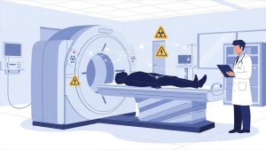
A radiology or imaging report is the written record of medical imaging examinations, such as MRI (Magnetic Resonance Imaging), CT (Computed Tomography), X-ray, ultrasound, PET (Positron Emission Tomography), and nuclear medicine tests. These reports are written by a radiologist, a physician who interprets your images and writes what they see. Most reports are shared with your doctor first, but many patients can now see them too.
Reading your imaging report can be hard because it uses medical terms you may not know. But with clear explanations, you can learn what the main parts of the report are and what standard terms mean. Learning this can help you communicate with your physician, better understand your results, and make informed decisions about your health.
In this article, you will learn how a radiology report is made, the main parts like Clinical indication, Findings section, and Impression section, and the key imaging terminology used in MRI, CT, X-ray, and ultrasound reports. We will explain terms so you can read your report with confidence.
Who Writes Your Imaging Report
Your imaging report is written by a radiologist, a physician trained to interpret images from various modalities, including MRI, CT, X-ray, and ultrasound. The radiologist reviews your photos and writes a structured report describing what is seen. This report is meant to help your doctor understand your scan results and plan your care.
Often, the report is first sent to the doctor who ordered the scan. That doctor then explains it to you. But with modern electronic health records, many patients can now access their own reports online. When you read your report, you may see clear medical terms that explain what the radiologist found.
Understanding who wrote your report and why it was written is the first step in learning how to read it.
Anatomy of an Imaging Report
Your imaging report has several main sections that help physicians interpret your scan results. These parts are built using structured radiology reporting, so each section has a clear purpose. A typical report often includes Clinical Indication, Technique, Comparison, Findings, and Impression sections. Each tells a piece of the story your images reveal.
Clinical Indication
The Clinical Indication section explains the rationale for ordering your imaging test. It shows the reason or clinical question your physician had when requesting the scan. This part helps the radiologist focus on the areas most related to your symptoms or medical concern.
For example, it may indicate that the imaging was performed for pain, injury, or to evaluate a known condition. This context is crucial because it guides the radiologist’s interpretation of your images. If the reason is vague, the radiologist may examine a broader area.
Technique
The Technique section describes how the imaging exam was done. It may include details of your imaging modality, such as MRI, CT, X-ray, ultrasound, or other modalities. It also notes whether a contrast agent was used.
This part is primarily for medical records and future reference. For example, it may state that the images were acquired with specific settings or that a contrast agent was administered to enhance the visibility of structures.
Comparison
The Comparison section lists any past imaging studies that the radiologist looked at to compare with your current images. Seeing changes over time can help doctors track progress or detect new issues.
If no prior imaging is available, this section may be omitted. Comparing with prior scans can be very helpful, especially if you are tracking a condition or follow-up after treatment.
Findings
The Findings section is the detailed part of your report. It lists the radiologist’s observations from the images. This might include descriptions of typical structures and any abnormalities, such as lesions, masses, nodules, or areas that differ from expected morphology.
The radiologist describes what was seen in objective language without yet interpreting its meaning. It often employs precise imaging terminology and indicates whether each area appears normal or abnormal.
Impression
The Impression section is the most critical part of the report. Here, the radiologist writes a summary of the key observations from the Findings, explains their likely interpretation, and answers the clinical question posed in the Clinical indication.
This section often gives a diagnostic assessment, suggests possible causes, and may include a differential diagnosis (a list of potential explanations). It may also recommend follow-up imaging or other next steps.
Understanding each section helps you read your imaging report with confidence. You can see what was done, why it was done, what the radiologist saw, and what it means in clear terms before talking with your doctor.
Standard Terms You’ll See & What They Mean
When you read an imaging report, you will see many key imaging terminology words that describe what the radiologist observed in your scan. These terms appear most frequently in the Findings and Impression sections and help present clinical imaging findings clearly and accurately. A good way to understand your report is to learn what common phrases and words mean in simple language.
General Report Words
- Normal/ unremarkable indicates that nothing unusual was observed in that area of the body.
- An incidental finding is a result that was not related to the reason the scan was performed but was discovered unexpectedly.
- A follow-up is recommended, indicating that the radiologist recommends another imaging test later to assess changes over time.
- Assessment/Impression is a summary that explains what the radiologist believes the images show. This is often the most critical part of your report.
- Comparison refers to prior scans that the radiologist reviewed to determine whether anything has changed.
Words That Describe What Was Seen
These terms describe what the radiologist saw on the images:
- A lesion is an area that differs from normal tissue.
- A nodule is a small, round lesion seen on imaging.
- Mass is a larger abnormal area that may require further testing.
- An artifact is an image feature that is not part of the body and arises from technical factors.
- Opacity/Radiopacity refers to an area that looks white or dense on an X-ray or CT image.
- Radiolucent means an area looks dark or less dense on imaging.
Helpful Words by Imaging Type
- In X-ray imaging and CT scans, terms such as attenuation and radiodensity describe the degree to which tissue blocks X-rays.
- In MRI, terms such as signal intensity indicate whether tissue appears bright or dark in certain views.
- In Ultrasound scans, terms such as echogenicity and hypoechoic describe how sound waves reflect from tissues.
- Contrast agents, or contrast dyes, are substances used in MRI or CT to enhance visualization of blood vessels or organs.
- Hounsfield units are a unit used to quantify tissue density in CT scans.
Standard Systems You Might See
- BI-RADS is a system used to describe breast imaging results, report risk levels, and provide a standardized format for reporting.
- LI-RADS is a way to describe liver imaging results, especially if cancer is possible.
- The RadLex radiology lexicon is a standard list of terms used by radiologists to describe imaging findings consistently.
Learning these standard terms can make your imaging report easier to read and help you talk more clearly with your doctor. Ask your provider if any words are still unclear so you fully understand your health information.
Modality-Specific Insights
In an imaging report, you may see terms that depend on the imaging modality used. Each type of scan uses different technology to create pictures of the inside of your body, so the key imaging terminology explains what the radiologist saw and how they saw it. Understanding these terms can help you read your report more clearly.
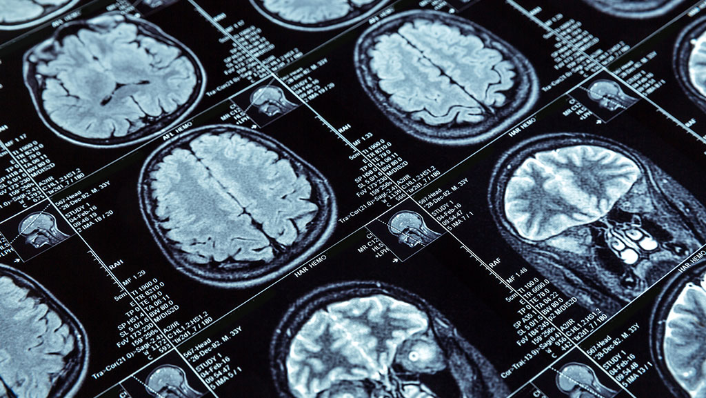
MRI (Magnetic Resonance Imaging)
In an MRI report, the radiologist describes how tissues show up using magnetic fields and radio waves. The words often focus on how bright or dark areas appear and whether a contrast agent was used. If the report mentions an artifact, it indicates an image feature that is not part of your body and originates from the scanning process (e.g., motion or signal artifacts).
MRI often includes details like:
- Whether contrast was given and how tissues were enhanced
- Bright or dark areas on signal intensity
- Findings related to organs, discs, or soft tissues
This helps physicians determine whether areas are normal or may require further study.
CT Scan (Computed Tomography)
A CT scan report uses X-rays to make cross-sectional pictures of your body. It measures how dense tissues are. A CT report may talk about:
- Areas that are more or less dense
- How structures look compared with normal tissue
- Any abnormal signs like nodules, lesions, or areas of increased opacity
These details help your physician assess whether there are changes in organs, bones, or other tissues.
X-ray Imaging
In X-ray reports, terms indicate the degree to which tissue blocks X-rays (radiopacity) or allows them to pass through (radiolucency). Dense structures, such as bones, appear bright, whereas air seems dark. You may find related descriptive terms in the Findings section that explain what the radiologist observed.
Ultrasound Scan
Ultrasound uses sound waves to make pictures. A report will use terms such as echogenicity to indicate how healthy tissues reflect sound. For example:
- Hypoechoic means an area makes fewer echoes and appears darker
- Hyperechoic refers to an area that produces more echoes and appears brighter.
These terms help describe tissues and possible abnormalities.
Standard Reporting Systems You Might See
Some imaging reports use standardized systems to make results easier to compare and understand:
- BI-RADS is used in breast imaging to communicate risk levels and guide follow-up care.
- Many reports are written in standard medical terminology, using sources such as the RadLex radiology lexicon, which helps radiologists use consistent terms when describing findings. rsna.org
Learning how each type of imaging works and what the key terms mean can help you read your Imaging report with more confidence before discussing results with your doctor.
Example: How to Read a Sample Report
Here is a simple example to help you learn how real imaging reports are written and how you can read them. In this example, you will see standard imaging report sections like Clinical indication, Technique, Comparison, Findings, and Impression. Real reports follow this structure, so it is easier for doctors to share information clearly.
Sample Report
Patient Information
Name, date of birth, and the date and type of the exam (e.g., MRI (Magnetic Resonance Imaging) or CT scan (Computed Tomography)) appear at the top of the report.
Clinical Indication
This part explains the reason your doctor ordered the scan. It presents the clinical question the radiologist should answer based on the images. For example, if you had back pain or a possible injury, the radiologist would focus on that area.
Technique
The Technique section tells how the imaging study was done. It lists the imaging modality, such as X-ray, ultrasound, MRI, or PET(Positron Emission Tomography). If a contrast agent was used, it is noted here as well.
Comparison
If you had a previous scan, the radiologist will compare today’s images to the older ones. This shows temporal changes and helps clinicians assess progress or new findings.
Findings
In this section, the radiologist writes what they saw in the images. They describe the clinical imaging findings, including normal areas and things like lesions or nodules. The radiologist may note whether an image is unremarkable (regular) or whether an artifact (a technical error in the image) is present.
Impression
The Impression is the radiologist’s summary of the most important results. It answers the clinical question and may suggest causes or next steps, such as follow-up tests. This is usually the section your doctor pays most attention to when deciding treatment.
What to Do if You’re Confused by Your Report

Reading imaging reports, including MRI (Magnetic Resonance Imaging), CT (Computed Tomography), X-ray, ultrasound, PET (Positron Emission Tomography), or nuclear medicine, can be challenging. Many patients find medical terminology confusing and may feel anxious about the results. Studies show that many patients feel confused or frightened when they attempt to read their radiology reports independently because the reports are written in medical terminology for physicians, not patients. About 85 percent of patients in one study reported confusion about medical terms in their imaging results.
Talk With Your Doctor First
Your first step should be to ask your doctor (the provider who ordered the test) to explain the report in plain words. This doctor knows your symptoms and medical history and can connect the clinical imaging findings in your report to your health in a simple way. If the report mentions an incidental imaging finding that was not related to the reason you had the test, your physician can help explain whether it matters and what next steps might be.
Ask for a Radiologist Review
If your doctor still has questions, they can contact the radiologist who wrote the imaging report. Many imaging centers will allow your doctor or you to speak with a radiologist to review the imaging report terminology more clearly. RadiologyInfo.org offers articles and videos designed to explain imaging reports in plain language and help you understand sections such as Findings and Impression.
Electronic Access and Patient Resources
Many patients can access their radiology reports and images online via electronic health records or patient portals. Reading your report ahead of your follow-up appointment can give you more time to prepare questions and understand what your provider will discuss with you.
Get a Second Opinion if Needed
If the imaging report is still unclear or if you and your doctor disagree with the findings, consider a second opinion from another radiologist. This can be especially helpful if you are being advised to undergo surgery based on imaging or if the report appears vague or confusing.
By talking with your doctor, using reliable patient resources, and asking for help when needed, you can make reading your imaging report easier and less stressful.
Frequently Asked Questions
Here are common questions people have when they read their imaging report, along with clear answers based on trusted medical sources. These FAQs help you understand your report better and make it easier to talk with your doctor.
What does a radiology or imaging report show?
A radiology report is a written summary of what a radiologist saw in your medical imaging test, such as an MRI (Magnetic Resonance Imaging), CT scan (Computed Tomography), X-ray imaging, Ultrasound scan, PET scan (Positron Emission Tomography), or nuclear medicine exam. It includes details such as the type of exam, the rationale for its administration, the findings, and an overall interpretation.
Why is my imaging report hard to understand?
Radiology reports are written mainly for doctors, so they use key imaging terminology that may be new to you. Seeing medical words and detailed descriptions can feel confusing if you aren’t familiar with terms like the Findings section, the Impression section, or clinical indication.
Why does my report compare old and new scans?
Your report may include a Comparison section that looks at your current images and any previous ones. This allows the radiologist to assess whether changes have occurred over time, which can help track disease progression or healing.
What does the Impression section mean?
The Impression section of the report summarizes the most important results from the Findings section and presents the radiologist’s overall interpretation. It often suggests what the observations might mean and may recommend follow-up care if needed.
Does “Unremarkable” mean serious?
In radiology, unremarkable usually means that no significant abnormality was seen in that area. This does not imply that something is wrong. This term is often used to show a normal appearance.
What if I see an “Incidental finding”?
An incidental finding is something that was not related to the reason you had the test but was found anyway. Your doctor can explain whether it needs follow-up or if it is harmless.
Can I access my imaging report online?
Yes. Many patients can view their own imaging reports and images through online Electronic Health Records or patient portals. This can help you prepare questions for your doctor before your follow-up appointment.
What should I do after reading my report?
Consult the doctor who ordered your test to review the results in clear language. They can explain how findings relate to your symptoms and help plan next steps. If you remain unsure, you may request to speak with the radiologist or seek a second opinion.
Does the report list possible diagnoses?
Sometimes the Impression section may include a differential diagnosis, a list of possible causes for the findings when a single diagnosis is not definitive. Your doctor will help you understand what these possibilities mean for your care.
Conclusion
Understanding your imaging report helps you take an active role in your health. A radiologist writes a radiology report and includes sections such as Clinical Indication, Technique, Comparison, Findings, and Impression. Each part clearly explains what your MRI (Magnetic Resonance Imaging), CT scan (Computed Tomography), X-ray, ultrasound, PET scan (Positron Emission Tomography), or nuclear medicine test shows and what it means for your care. The Impression section, or conclusion, is a key component because it summarizes the most critical points from the detailed observations in the Findings section and may suggest next steps or follow-up care.
Reading your report with confidence means knowing what each section shows and asking your physician about any confusing terms. Many imaging centers and health systems now allow patients to access their imaging reports and images through Electronic Health Records or patient portals. This can give you time to look up terms and prepare questions before your follow-up appointment.
The purpose of the radiology report is to help your healthcare team make the best decisions about your diagnosis and treatment. It acts as a formal consultation from the radiologist to your doctor and plays a vital role in your ongoing care. Learning to interpret key terms and understand the meaning of the impression can make your subsequent conversation with your provider more precise and more useful for your health.