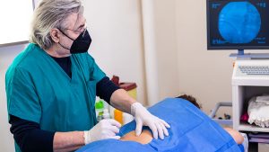
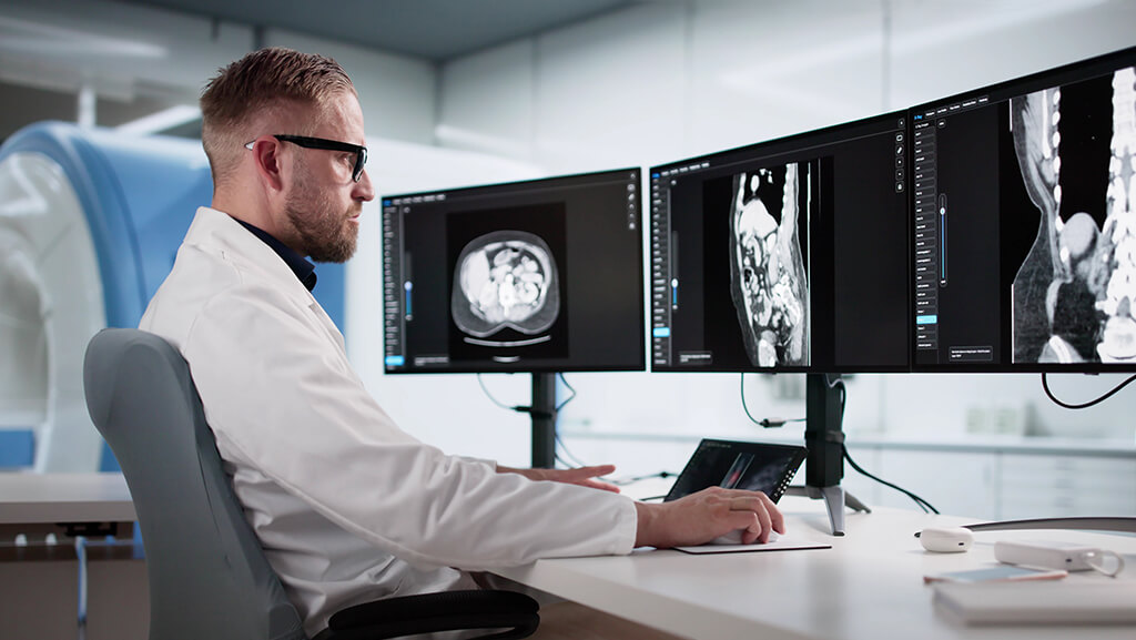
Imaging is a big part of modern neurology. It lets doctors look inside the brain, spinal cord, and nerves without surgery. This helps them identify the cause of symptoms such as weakness, seizures, memory loss, headaches, or sudden speech difficulties. Imaging can reveal structural, functional, and molecular changes in the brain. These views make diagnosis faster and more exact.
Doctors use neuroimaging in neurology to answer key questions. Is there bleeding or a stroke? Is a brain tumor present? Are there signs of neurodegeneration or demyelinating disease? Imaging also supports neuroradiology diagnostics and helps with clinical decision support for neuroimaging. It does not replace the doctor’s exam, but it adds strong proof.
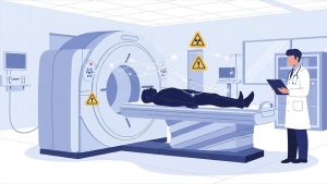
Different tests are used for different needs. Computed Tomography (CT) is fast and widely used in emergencies. Magnetic Resonance Imaging (MRI) gives more detail and is used for many long-term problems. Positron Emission Tomography (PET) and Single-Photon Emission CT (SPECT) show how the brain works, not just how it looks. Ultrasound (Carotid & Transcranial Doppler) checks blood flow in head and neck vessels. Together, these tools form multimodal brain imaging for better care.
Imaging can also act as a set of imaging biomarkers in neurology. These biomarkers help doctors detect disease early, track change, and guide treatment. This is important because many diseases start years before clear symptoms appear. Imaging helps spot those early changes and supports early care.
Core Neuroimaging Modalities and What Each Reveals
Doctors choose an imaging test based on the patient’s symptoms and how fast they need answers. Some tests show brain structure. Others show blood flow or brain activity. Using the right test helps doctors find the cause of a problem sooner.
MRI
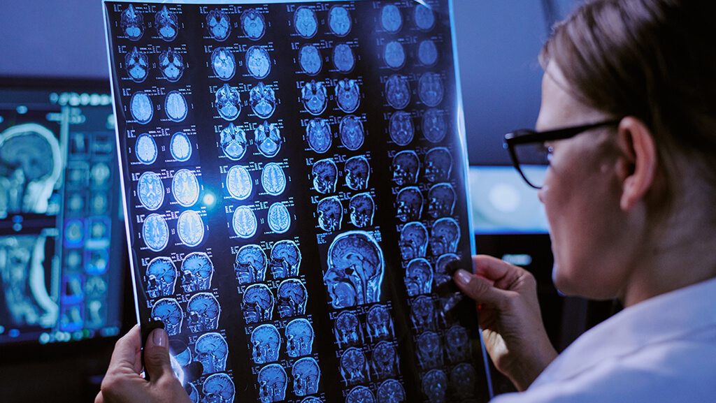
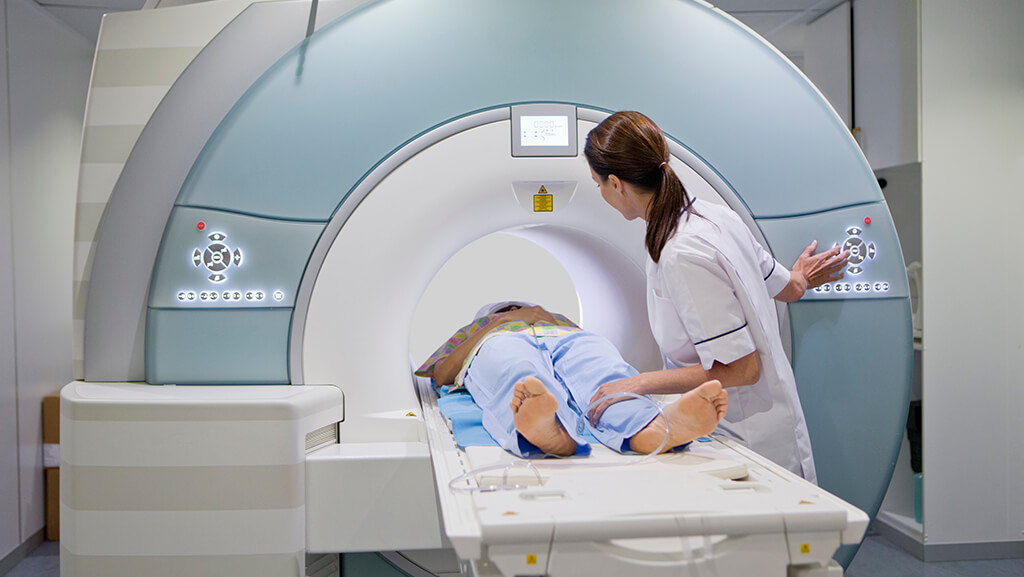
MRI is one of the most useful tools in neurology. It uses strong magnets, not radiation. This makes it safe for many people. MRI gives very clear pictures of soft brain tissue. It helps doctors visualize the white matter, gray matter, brainstem, and spinal cord.
MRI can use different scan types, called sequences. Each sequence shows a different kind of change. For example, diffusion scans can spot early stroke, and SWI scans can find tiny bleeds. With contrast dye, MRI can also show active inflammation or tumors. Because of this, MRI is a top choice for many long-term brain and nerve diseases.
CT
CT is fast and widely available. A CT scan uses X-rays, so it exposes you to some radiation. But it is very helpful in emergencies. Doctors often pick CT first when a person has sudden weakness, a head injury, or signs of bleeding. CT is strong for finding intracerebral hemorrhage, skull fractures, large tumors, or swelling that raises brain pressure.
CT can be paired with specialized tests such as CT angiography and CT perfusion. These show blood vessels and blood flow. They help doctors check for vessel blockage, aneurysm, or brain areas at risk during an acute stroke.
PET
PET shows how the brain works, not just how it looks. It uses a small amount of radioactive tracer. The tracer highlights areas of brain activity or disease proteins. FDG PET measures brain sugar use. Low sugar use in certain patterns can help diagnose types of dementia.
PET can also look for disease markers. Amyloid PET and tau PET help confirm Alzheimer’s type changes in people with memory problems. New guidelines explain when these PET scans are most useful for diagnosis and care planning.
SPECT
SPECT is another functional scan. Like PET, it uses a tracer. SPECT often measures brain perfusion, which refers to the flow of blood through brain tissue. It can support diagnosis in dementia, epilepsy, and some blood flow disorders. It is often more available and cheaper than PET, though it gives less detail.
Ultrasound
Ultrasound uses sound waves, not radiation. In neurology, it is mainly used to study blood vessels. A carotid ultrasound looks for narrowing in neck arteries. Transcranial Doppler checks flow in major brain arteries. These tests help monitor stroke risk and changes in blood flow over time.
Advanced Imaging Techniques That Improve Diagnostic Precision
Basic scans like MRI and CT are very helpful. But doctors often need more detail. Advanced imaging adds that detail. It can show very early damage, tiny blood flow changes, and hidden white matter injury. This helps doctors make a faster, more accurate diagnosis.
Diffusion-Weighted Imaging (DWI)
Diffusion-weighted imaging (DWI) is a special MRI method. It shows how water moves in brain tissue. When a person has an Acute ischemic stroke, the movement of water changes right away. DWI can find a stroke within minutes to about an hour, much earlier than standard MRI or CT. Because of this, it is a core tool in MRI vs. CT for stroke workup and emergency care.
Doctors also use DWI in other problems. It helps tell a brain abscess from a tumor and helps spot acute infection changes. This makes diagnostic imaging of brain disorders more reliable.
Perfusion Imaging
Perfusion imaging shows how blood flows through the brain. It is widely used in stroke care. CT perfusion in acute stroke and MR perfusion techniques help doctors see two zones:
- the infarct core, which is already damaged
- the penumbra, which is hurt but can still be saved
Finding the penumbra guides urgent treatment and improves results. That is why perfusion is part of modern neurovascular imaging and clinical decision support for neuroimaging.
Perfusion tools also help with tumors. Perfusion MRI tumor grading shows how much blood a tumor uses. This supports glioma imaging features and treatment planning.
Diffusion Tensor Imaging (DTI) and Tractography
Diffusion tensor imaging (DTI) is a more detailed form of diffusion MRI. It maps white matter tracks. With tractography in white-matter disease, doctors can see if brain pathways are damaged or pushed aside.
This is useful in:
- imaging traumatic brain injury (TBI) and diffuse axonal injury MRI
- imaging for multiple sclerosis lesions and other demyelinating disease MRI
- surgery planning near important brain areas
DTI can show hidden injury in Traumatic brain injury (TBI), even when the CT looks normal. It also helps predict recovery.
Functional MRI (fMRI)
Functional MRI (fMRI) shows which brain areas are active during tasks like speaking or moving. It is used for fMRI brain activation mapping before surgery. Doctors also use resting-state fMRI connectivity to map brain networks without hard tasks.
This helps in:
- Presurgical epilepsy imaging and seizure focus localization
- Brain tumor MRI protocol before removal
- Protecting speech, movement, and vision areas in surgery
Because it is noninvasive, fMRI is now common in planning safe brain operations.
MR Spectroscopy (MRS)
Magnetic Resonance Spectroscopy (MRS) is another MRI add-on. It measures brain chemicals. MR spectroscopy (MRS) helps doctors study tissue that looks unclear on normal scans.
It supports:
- spectroscopy for tumor characterization
- infection workups like encephalitis, neuroimaging
- checking for active inflammation
This helps improve neuroradiology diagnostics and reduces the risk of incorrect diagnoses.
Imaging Across Major Neurological Disease Categories
Imaging helps doctors answer one main question. What disease is causing these signs? Below are the most common disease groups and how imaging helps in each one.
Cerebrovascular disease: stroke, aneurysm, AVM
When a person may have a stroke, time is very important. Doctors often start with a non-contrast CT. This quick scan helps rule out intracerebral hemorrhage. If there is no bleed, they look for a blocked vessel.
Next, CT angiography (CTA) or MR angiography (MRA) can show the brain arteries. These scans help detect large-vessel occlusion, cerebral aneurysms, or arteriovenous malformations (AVMs).
For early brain injury, diffusion-weighted imaging (DWI) on MRI is highly sensitive. Perfusion imaging, like CT perfusion (CTP) or MR perfusion, shows the penumbra, the brain area that may still be saved. This guides clot removal or clot-busting treatment.
Neurodegenerative disease: Alzheimer’s, Parkinson’s, FTD
In memory or movement problems, imaging looks for slow changes over time. Structural MRI can reveal patterns of brain atrophy, such as hippocampal atrophy in Alzheimer’s disease.
FDG PET shows brain sugar use. Low use in certain areas helps tell Alzheimer’s disease, frontotemporal dementia (FTD), and vascular dementia apart.
Doctors may also use amyloid PET and tau PET. These scans look for disease proteins. They help confirm Alzheimer-type changes, especially when the diagnosis is unclear.
For Parkinson’s disease and other movement disorders, doctors may use dopamine scans like DaT imaging. These show early changes in the brain’s dopamine system.
Demyelinating and autoimmune disease: MS and related disorders
In multiple sclerosis, MRI is the main test. Doctors look for lesions in typical locations, such as around the ventricles, brainstem, and spinal cord. They also look for new lesions on later scans. This shows disease spread in space and time.
Gadolinium enhancement can show active inflammation. Follow-up MRI helps track activity and treatment response.
Brain tumors and neuro oncology
For suspected tumors, contrast-enhanced MRI is the key scan. It shows the size, location, swelling, and whether the tumor is breaking the blood-brain barrier.
Advanced tools add more detail. Perfusion MRI helps estimate tumor grade by showing blood supply. MR spectroscopy (MRS) can check tumor chemicals. DTI tractography maps white-matter pathways, so surgeons can avoid vital areas.
Imaging is also used after treatment to see if a tumor is shrinking or growing again.
Epilepsy
Many people with repeated seizures need imaging. A high-quality MRI can find scars, small malformations, or tumors that may cause seizures. This is a core part of epilepsy diagnosis and care.
If surgery is planned, doctors may order additional tests. PET and SPECT can show low-function areas between seizures. fMRI and DTI help map speech and movement areas, making surgery safer.
Traumatic brain injury
After a head trauma, doctors usually start with a CT. It quickly finds bleeding, skull fractures, or swelling.
If symptoms persist despite a normal CT, an MRI can help. SWI can show small bleeds, and DTI can show diffuse axonal injury (DAI), which is tiny damage to white matter tracks.
CNS infections and inflammation
In infections like meningitis or encephalitis, MRI shows patterns of swelling and tissue injury.
DWI helps detect pus in a brain abscess and differentiate an abscess from a tumor. MRS can add chemical clues when the scan is unclear.
Imaging also tracks problems like hydrocephalus or stroke caused by infection.
How Clinicians Choose the Right Imaging Test
Doctors do not pick a scan at random. They follow symptoms, risk signs, and trusted guidelines. The goal is to choose the test that gives the clearest answer with the least delay. Large professional groups like the American College of Radiology (ACR) and the American Academy of Neurology (AAN) publish imaging guidance for many neurologic conditions. Stroke care is also guided by the American Heart Association/American Stroke Association (AHA/ASA), and neuroradiology standards are set by groups such as the American Society of Neuroradiology (ASNR).
When symptoms are sudden and severe
If a person has signs of acute stroke or brain bleeding, doctors need speed. They often start with a non-contrast CT. This helps rule out intracerebral hemorrhage very fast. After that, they may add CT angiography (CTA) to look for a blocked vessel and may use CT perfusion (CTP) to see brain tissue that can still be saved. These steps align with stroke imaging guidance from the AHA/ASA and RSNA stroke statements.
When seizures happen
For a first seizure or repeated seizures, doctors usually choose MRI because it shows small scars, tumors, or malformations better than CT. If surgery may be needed, they may add a PET or SPECT scan to identify the active problem area. This plan follows common imaging paths in epilepsy care.
When headaches have warning signs
Most headaches do not need a scan. But imaging is advised when red flags are present. Red flags include new severe headache, head injury, cancer history, weak immune system, or new problems like weakness or confusion. When imaging is needed, MRI is often preferred because it finds more causes than CT, except in emergency cases. These points are supported by ACR headache criteria and patient-focused neurology guidance.
When memory or thinking slowly gets worse
If a person has slow memory loss or confusion, doctors often start with an MRI. They look for patterns of brain atrophy, old strokes, or tumors. If the cause is still unclear, they may suggest FDG PET, amyloid PET, or tau PET to help tell which type of dementia is present. This helps separate Alzheimer’s disease, frontotemporal dementia, and vascular dementia.
When trauma happens
After a head injury, doctors start with a CT because it is quick and good for bleeding and fractures. If symptoms persist and the CT is normal, they proceed to MRI for more detailed evaluation, especially to assess for hidden injury.
Other practical factors
Doctors also think about these points before ordering a scan:
- Speed and access. CT is faster and more available in many hospitals.
- Radiation. CT, PET, and SPECT use radiation, so doctors avoid them when not needed, especially in children.
- Metal or implants. Some people cannot have an MRI.
- Contrast safety. Some scans need dye. Doctors check kidney health and allergy risk first.
- Cost and follow-up. MRI and PET may cost more, so they are used when they add real value.
Choosing the right test helps avoid wasted scans and speeds up care. It also supports clinical decision support for neuroimaging in daily practice.
Benefits and Limitations of Neuroimaging

Neuroimaging provides doctors with powerful tools to diagnose brain and nerve conditions. But every scan has limits. Knowing both sides helps patients and doctors make good choices.
Main benefits
- Fast and clear diagnosis
Imaging can show bleeding, a blocked vessel, swelling, or a mass very quickly. This is lifesaving in emergencies like acute stroke or head injury.
- Early detection
Some diseases start slowly. Imaging can find early changes before symptoms get severe. For example, MRI can detect early tissue damage, and PET can detect early changes in brain function.
- Better disease typing
Many neurological diseases look similar at first. Imaging patterns help doctors tell them apart. This helps with the right treatment plan.
- Guides treatment and surgery
Imaging shows where a lesion is and its size. Advanced scans can map vital brain areas before surgery. This lowers risk.
- Tracks change over time
Doctors use follow-up scans to see whether a disease is worsening or whether treatment is working. This is common in stroke recovery, tumors, and immune diseases.
Main limitations
- Incidental findings
Scans can show spots that are not causing symptoms. These are called incidental findings. Most are harmless, but they can cause stress and lead to extra tests. Studies show they are common on brain MRI.
- False positives and false negatives
Imaging is powerful, but not perfect. A scan may appear abnormal when the brain is fine, or normal when the disease is early or small. This is why imaging must be interpreted in the context of the medical exam and history.
- Radiation exposure
CT, PET, and SPECT use ionizing radiation. The dose is usually small, but doctors try to avoid extra scans, especially in children and pregnant patients.
- Contrast risks
Some MRI scans use gadolinium contrast to show inflammation, tumors, or blood vessel leaks. The FDA and major safety groups note that tiny amounts of gadolinium can stay in the body, so doctors use contrast only when it adds clear value. People with kidney disease need special care.
- Cost and access
MRI and PET can be expensive and may not be available everywhere. This can delay diagnosis for some patients.
- Not a full answer by itself
Imaging shows structure or function, but it does not always explain the full cause. Blood tests, spinal fluid tests, and the doctor’s exam often remain needed.
Emerging Trends Shaping the Future
Brain imaging is improving fast. New tools help doctors see disease earlier and more clearly. These changes may make diagnosis quicker and more personal for each patient.
1. AI and deep learning in neuroimaging
AI in neuroimaging diagnosis means computers help read scans. Deep learning image analysis can spot patterns that are hard for people to see.
Today, AI is being used to:
- Find early stroke changes on CT or MRI
- Help with lesion segmentation in MRI
- Support faster scan reading in busy hospitals
- Improve detection of brain tumors, trauma, and dementia patterns
AI does not replace doctors. Instead, it acts as a smart helper, speeding up care and reducing missed findings.
2. Radiomics and quantitative imaging
Radiomics in brain disease converts scan images into numerical data. These numbers describe shape, texture, and signal strength. When combined with AI, radiomics can improve diagnosis and predict how fast a disease may change.
New work shows that radiomics helps in the diagnosis of Alzheimer’s disease and other dementias. It may also help in tumors by tracking how a mass responds to therapy.
3. Quantitative MRI biomarkers
A regular MRI gives pictures. Quantitative MRI adds exact measurements. Examples include values for tissue water, iron, blood flow, and brain volume.
Researchers are building shared standards so these measures can be compared across hospitals. This strengthens the utility of imaging biomarkers in neurology for precision medicine.
Over time, quantitative MRI may help doctors:
- Detect very early neurodegeneration
- Measure disease growth more precisely
- Check if treatment is working
4. Better protein imaging with PET
PET is improving for dementia care. Amyloid PET and tau PET imaging can detect Alzheimer-related proteins in living patients.
In 2025, expert groups updated rules for when these scans are most helpful. The goal is to use PET in the right patients, such as those with unclear memory decline or mixed symptoms. This improves scan value and avoids overuse.
5. Hybrid PET MRI and multi-scan care
Some centers now use hybrid PET MRI. This combines PET functional data with MRI structural data in a single session. It can show both where brain tissue is changing and how it is working.
Hybrid scans may help in:
- Dementia subtyping
- Difficult tumor cases
- Complex epilepsy workups
This supports a future in which multimodal brain imaging becomes the standard for diagnosing hard cases.
Practical Takeaways
Here are simple points to remember about the role of imaging in diagnosing neurological diseases. These ideas align with major reviews and guidelines on how scans help in real care.
1. Imaging helps find the cause fast
Doctors use imaging to determine whether symptoms are caused by bleeding, blocked blood flow, swelling, infection, or a mass. This is why imaging is crucial in sudden problems like stroke or head injury.
2. MRI and CT have different strengths
CT is fast and great for emergencies, like checking for brain bleeding.
MRI takes longer but shows more detail for long-term diseases, small lesions, and spinal cord problems.
3. Advanced MRI can show disease earlier
Special MRI methods such as DWI, perfusion, DTI, fMRI, and MRS provide additional detail. They help detect early stroke, hidden white matter injury, tumor type, and brain network changes.
4. PET and SPECT show how the brain works
PET and SPECT can reveal brain metabolism or blood flow. This helps in dementia and epilepsy cases where structure alone is not enough.
5. Dementia imaging is becoming more exact
MRI shows brain shrinkage patterns.
Newer PET scans can look for amyloid and tau proteins. Updated use criteria say these scans are most helpful when memory decline is unclear or mixed, and when results will change care.
6. Imaging guides treatment and follow-up
Scans help plan surgery, choose stroke treatment, and track if the disease is getting better or worse. Imaging biomarkers are now a key part of monitoring many neurologic diseases.
7. The right scan depends on symptoms
Doctors follow symptom-based pathways and guidelines to decide which test to use first. This lowers risk, saves time, and avoids extra scans.
8. Imaging is powerful but not perfect
Scans can miss early disease or show harmless findings. CT and nuclear scans use radiation, and contrast dye is used only when it adds real value. Imaging is best used in conjunction with the doctor’s exam and other tests.
FAQs about imaging in neurological diseases
1. What is the most accurate imaging test for neurological diseases
There is no single best test for every case. MRI is often the most detailed scan for many brain and spine problems because it shows soft tissue clearly. CT is best when doctors need a quick answer, such as in emergency care. PET and SPECT are used when doctors need to see how the brain works, not only how it looks. The right test depends on the disease and symptoms.
2. When is CT preferred over MRI for brain problems
Doctors choose CT first when time is critical. CT is common for suspected acute stroke, head injury, or possible brain bleeding. Stroke guidelines say that rapid CT helps rule out bleeding and can be followed by vessel and perfusion scans when needed.
3. How does PET help diagnose Alzheimer’s or Parkinson’s
PET can show brain metabolism and disease proteins. In patients with memory loss, amyloid PET and tau PET can help confirm Alzheimer’s type changes in the right patients. In movement problems, dopamine PET or similar scans can support diagnosis when symptoms are unclear.
4. Can MRI detect early stroke or multiple sclerosis
Yes. In stroke, a special MRI scan called DWI can detect brain injury very early, often within minutes. In multiple sclerosis, MRI shows typical lesion patterns and helps track new or active lesions over time. This is why MRI is the key imaging tool for MS diagnosis and follow-up.
5. What are advanced MRI techniques, like DTI or fMRI, used for
DTI maps white matter pathways and can show hidden injury in trauma or demyelinating disease. It is also used to plan safe surgery. fMRI shows brain activity during tasks or at rest, helping doctors map speech, movement, and vision areas before tumor or epilepsy surgery.
6. Are brain scans safe? What about radiation and contrast
Most brain scans are safe. MRI uses no radiation. CT, PET, and SPECT do use radiation, so doctors avoid doing them unless they are needed. Some MRI scans use contrast dye to show inflammation or tumors. Doctors first check kidney health and allergy risk. Safety guidance recommends using contrast only when it adds clear clinical value.
7. How do doctors decide which imaging test you need
Doctors follow symptom-based pathways and guidelines. For example, headache scans are usually done only when there are red flags like neurologic deficit, cancer history, immune problems, or trauma. In epilepsy, expert guidance recommends MRI for most people with repeated seizures and a dedicated epilepsy MRI protocol when needed. In stroke, guidance supports rapid CT first, then more tests if required.