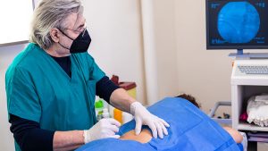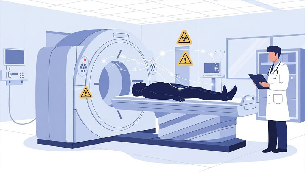
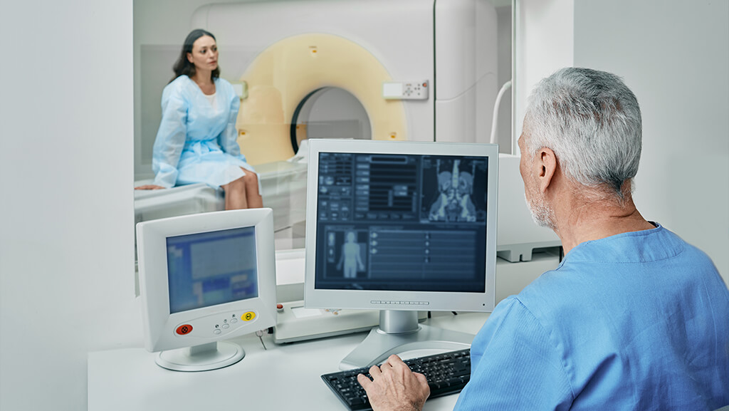
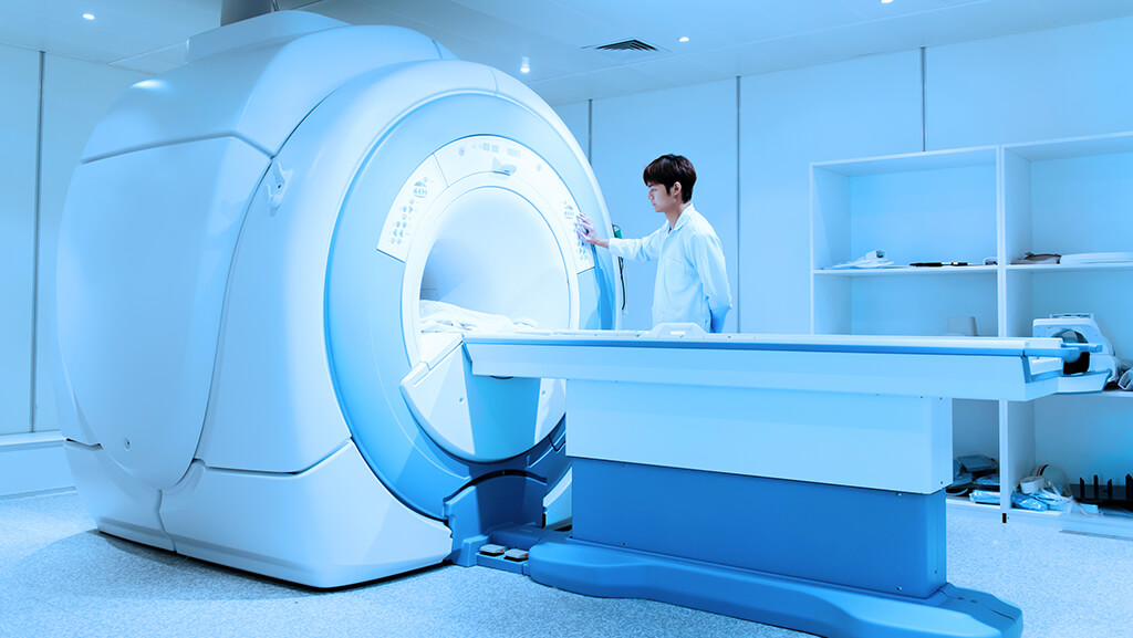
A CT scan, also called computed tomography, is a medical test that uses ionizing radiation to produce clear images of the inside of the body. Doctors use it in the radiology department because it is fast and can detect serious problems such as bleeding, injury, infection, or cancer. For many people, a CT scan can be lifesaving.
Still, many users search for information on radiation risks from CT scans to determine whether a scan is safe. CT uses X-rays, so it gives a radiation dose. This dose is often referred to as the effective dose in mSv (millisieverts). The dose is not the same for every scan. It depends on the body part, the scan plan, and the machine.
Radiation from CT can cause two types of effects. The first type is deterministic effects, which happen only at very high doses and are not expected in normal diagnostic CT. The second type is stochastic effects, which means a small chance of cancer that can increase with a higher dose. Scientists study this using models such as the BEIR VII risk model and the linear no-threshold model. These models help estimate lifetime attributable risk.
For most adults, the risk from one scan is small. But the risk can be higher in pediatric CT because kids are more sensitive and have more years ahead of them. Large studies have found a dose link with some cancers, like childhood leukemia after CT and brain tumor risk after CT, though the overall chance is still low for a single scan.
The good news is that modern CT care follows the ALARA principle, which means the dose should be as low as reasonably achievable. Tools like automatic exposure control, tube current modulation, and iterative reconstruction help reduce dose while maintaining high image quality. Many hospitals also follow diagnostic reference levels (DRLs) to avoid extra exposure.
1. What Is a CT Scan and Why Does It Use Radiation?
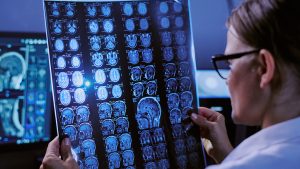
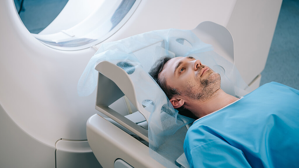
A CT (computed tomography) scan is a type of X-ray test. It takes many pictures from different angles. A computer combines them to create slices, like body views. These views help doctors see problems that a normal X-ray may miss.
CT uses ionizing radiation, which is the same type of energy used in other medical imaging tests that rely on X-rays. This is why people ask about CT scan radiation risk and computed tomography cancer risk. Radiation can damage cells. In rare cases, that damage may lead to radiation-induced malignancy years later. This is called a stochastic radiation effects risk. The chance is small for a single scan, but it can increase with a higher dose or with many scans.
Doctors still use CT scans a lot because the benefit-to-risk assessment often favors them. CT can be the fastest way to detect a serious problem, such as bleeding in the brain, a blood clot in the lung, or a hidden injury after a crash. In these cases, CT may save a life right away.
How CT Dose Is Measured
CT dose is usually described in a few ways.
- The CT dose index (CTDIvol) is the scanner’s output.
- Dose length product (DLP) combines the scan length and dose to indicate the total energy.
- size-specific dose estimate (SSDE) adjusts dose for body size.
- Doctors also talk about effective dose (mSv), which helps compare risk across scans.
A scan can be a head CT, a chest CT, or an abdominal CT. Doses vary a lot. A multiphase CT exam or CT angiography radiation test can deliver a higher dose than a single-phase scan.
Why Dose Can Change From One CT to Another
Your dose depends on the scan plan and machine settings.
- kVp selection impact and mA, and exposure time
- pitch and collimation effects
- scan length reduction
- Use of contrast-enhanced CT dose plans
- tube current modulation and automatic exposure control (AEC)
These tools help keep images clear while lowering the dose. New scanners also use iterative reconstruction and other dose-reduction technologies to reduce noise, thereby requiring less radiation.
Safety Ideas Doctors Follow
Radiology teams follow the ALARA principle. This means radiation should be as low as reasonably possible. They also use diagnostic reference levels (DRLs) and dose registries, such as the ACR Dose Index Registry, to compare and improve protocols. Groups such as the American College of Radiology (ACR), the Radiological Society of North America (RSNA), the Image Gently Alliance, and the Image Wisely campaign promote safe CT use, especially for kids.
What This Means for Patients
The key point is simple. CT uses radiation because it is an X-ray test. The radiation dose can slightly raise the lifetime attributable risk of cancer. For most adults, a single scan adds a very small risk. In pediatric CT, doctors take extra care because children are more sensitive to radiation. If you need a CT, the medical benefit is usually much bigger than the small radiation risk.
2. How Much Radiation Does a CT Scan Deliver
Many people ask, How much radiation does a CT scan give. The amount is called the radiation dose. Doctors often write it as effective dose (mSv). One mSv (millisievert) is a small unit of dose. A CT scan usually delivers a higher dose than a plain X-ray, but the dose can vary widely from one scan to another.
Typical CT Doses in Simple Numbers
These are common ranges for adults. Your dose may be lower or higher.
- The head CT dose is typically 1-2 mSv.
- The chest CT dose is typically 5-8 mSv.
- Abdominal CT dose and abdomen-pelvis CT dose are often about 8 to 15 mSv, and sometimes more.
- A multiphase CT exam of the abdomen can be much higher, even exceeding 20-30 mSv.
- Low-dose lung screening and lung cancer screening CT doses are lower, often around 1-2 mSv.
- Tests like coronary CT angiography risk or CT angiography radiation can be higher because they may need extra phases.
These numbers explain why people worry about CT scan radiation risk and computed tomography cancer risk. Still, the risk from one scan is usually small.
How CT Dose Is Measured
CT machines report several dose values.
- CT dose index (CTDIvol) shows the scanner output for that scan.
- Dose length product (DLP) combines the scan length with the dose, so it shows the total energy used.
- A size-specific dose estimate (SSDE) adjusts the dose for body size. A smaller body often needs less dose.
- These numbers help experts estimate the absorbed dose to the organ and the whole-body effective dose.
You may hear about CT dosimetry phantoms and Monte Carlo dose simulation. These are tools scientists use to test doses safely and improve CT dosimetry accuracy.
Comparing CT to Background Radiation
People also ask how CT compares to the radiation in daily life.
- We all get background radiation from nature every year.
- A head CT is like several months of background radiation.
- A chest CT is about two years of background radiation.
- An abdominal-pelvis CT can be like about three years of background radiation.
This comparison helps users understand risk in a real-world way.
Why Dose Can Vary So Much
Two people can get different doses from the same kind of CT.
Reasons include:
- Body size and shape, which affect SSDE.
- Scan plan, like contrast-enhanced CT dose and extra phases.
- Machine settings such as kVp, mA, exposure time, pitch, and collimation.
- Use of tube current modulation and automatic exposure control (AEC).
- Scan length and the amount of the body covered.
Studies show big dose differences across hospitals and scanners. This is why health groups track doses to improve care.
What This Means for Risk
Dose matters because cancer risk follows a dose-response relationship. Risk models such as the BEIR VII risk model and the linear no-threshold model estimate a small lifetime attributable risk from CT. These models are used by groups such as the International Commission on Radiological Protection (ICRP) and the National Academies.
For most adults, the risk per examination is low. The risk is higher in pediatric CT, so doctors work hard to keep the CT child-sized and follow ALARA.
3. What Risk Means in CT Scans
When people talk about radiation risks from CT, they usually mean a possible risk of cancer later in life. CT scans use ionizing radiation, so the body gets a small absorbed dose. Most of the time, this dose does not cause any quick harm. The worry is about effects that may happen many years later.
Two Main Types of Radiation Effects
Doctors and scientists split radiation effects into two groups.
Deterministic effects
- These occur only at high dose levels.
- The effect worsens as the dose increases.
- Examples include skin injury or lens damage in the eye, but these require very large doses.
- Normal diagnostic CT doses are far below these levels, so deterministic effects are not expected from routine scans.
Stochastic effects
- These happen by chance.
- The chance increases with the dose.
- The main stochastic concern is radiation-induced malignancy, meaning a cancer caused by radiation.
- This is why you may hear terms like stochastic radiation effects, radiation-induced malignancy, or dose response relationship in CT safety talks.
In simple terms, deterministic effects require a large dose and occur quickly. Stochastic effects can happen after small doses, but the chance is low.
How Scientists Estimate Cancer Risk
Because CT doses are low, doctors cannot immediately see a person’s cancer risk. So they use large studies and math models to estimate risk.
- The linear no-threshold model says that any amount of radiation may pose a small risk, and that risk rises in a straight line with dose.
- The BEIR VII risk model uses data from many groups to estimate cancer risk at low doses.
These models help estimate lifetime attributable risk. That means the extra chance of cancer over a whole life after a scan. The number is still small for one scan, but it matters more in children and in people who get many scans.
What Studies Show About Real World CT Risk
Large cohort studies in children found a clear dose-response relationship. Higher cumulative radiation dose from repeat scans was linked with a higher risk of childhood leukemia after CT and brain tumor risk CT. Even so, the absolute risk per scan remained low.
Newer studies in North America also support the idea that risk rises with higher cumulative dose, especially to bone marrow and the brain.
4. What Do Studies Say About Cancer Risk from CT
Researchers have studied CT for cancer risk for many years. They look at two things. First, they measure effective dose (mSv) from real scans. Second, they follow large groups of people to see if cancer happens more often after CT. Most results show the same pattern. The risk per examination is small, but it increases with higher doses and more scans over time.
4.1 Adults
For adults, most CT scans deliver doses far below the deterministic-effect thresholds. So the main worry is stochastic effects, meaning a small chance of cancer later. Risk models such as the BEIR VII risk model and the linear no-threshold model are used to estimate this risk. These models convert DLP, CTDIvol, and SSDE into risk estimates, such as lifetime attributable risk (LAR).
A large 2025 modeling study using U.S. data estimated that CT scans performed in one year could lead to many future cancers in the population. The study also found that, even though the risk per scan is higher in children, most projected cancers come from adults because adults get many more CT scans. The abdominal and pelvic CT doses and the chest CT dose contributed significantly, as these scans are common and can entail higher doses.
Risk in adults changes with:
- Radiation sensitivity by age
- Sex specific radiation risk
- Body area scanned, such as head CT dose, chest CT dose, or abdominal CT dose
- Use of multiphase CT exams or CT angiography radiation
- Cumulative radiation dose from repeat scans
Common cancers linked in models include lung, colon, leukemia, and bladder, but the added chance for one scan stays low. The key idea is to benefit first. If a CT is needed for trauma, stroke, or cancer care, the medical help is usually much larger than the small long-term risk.
4.2 Children and Teens
Studies show that pediatric CT risk is higher than adult risk. Children have faster-growing tissues and more years to live, so the same dose carries a greater lifetime risk. This is why groups like Image Gently Alliance push hard for low-dose CT protocols in kids.
The most well-known childhood study followed many children who had CT scans. It found a clear dose-response relationship. Higher dose to the brain was linked with a higher risk of brain tumors after CT, and higher dose to bone marrow was linked with a higher risk of childhood leukemia after CT. The increase was small in one scan, but it showed that risk does rise with increasing dose.
Other large reviews found similar results. Children exposed to CT had a higher later cancer rate than children without CT. Again, the absolute risk per scan remained low, but the pattern supports careful dose control.
Risk in kids depends on:
- Age at scan, with the highest risk in infants
- Scan region and organ absorbed dose
- Repeat CT scans and follow-up imaging
- Protocol choices like kVp selection impact and scan length reduction
5. Factors That Increase or Decrease CT Radiation Risk
The radiation risks from CT are not the same for everyone. Even two people who get the same test can receive different doses and face different risks. Doctors look at many parts of the scan to keep the risk low.
5.1 The Body Part Scanned
Radiation dose depends on where the CT is done.
- The head CT dose is usually lower than the body scans.
- The chest CT dose is higher because the scan covers more tissue.
- The abdominal CT dose and abdomen-pelvis CT dose can be higher still.
- Tests like CT angiography, radiation, or coronary CT angiography may use special timing that can raise the dose.
A higher dose increases the likelihood of stochastic radiation effects, such as a small radiation-induced malignancy risk.
5.2 Age and Sex
Your body’s response to radiation depends on age and sex.
- Radiation sensitivity increases with age.
- The pediatric CT risk is higher because kids have growing tissues and longer lives ahead.
- Sex-specific radiation risk can matter, too. For some cancers, women may have a slightly higher risk after the same dose.
This is why doctors use child-sized protocols for kids and follow Image Gently Alliance goals.
5.3 Body Size and Patient Conditions
Body size changes the needed dose.
- A larger body may need more radiation to get a clear image.
- A smaller body often needs less.
- This is why experts use size size-specific dose estimate (SSDE) to match the dose to the patient.
Certain health needs also matter. A very sick patient may need a longer or repeat scan, which can increase the dose.
5.4 Scan Settings and Protocol Choices
CT dose depends a lot on scanner settings.
Important settings include:
- kVp selection impact
- mA and exposure time
- pitch and collimation effects
- scan length reduction
- multiphase CT exams
- contrast-enhanced CT dose plans
A larger scan area or additional phases can increase the CT dose index (CTDIvol) and the dose-length product (DLP).
5.5 Dose Saving Technology
Modern CT machines have tools to lower the dose.
- Automatic exposure control (AEC) changes the dose in real time.
- Tube current modulation reduces the dose when less is needed.
- Iterative reconstruction and newer smart reconstruction let doctors reduce the dose while keeping images sharp.
These tools help hospitals follow the ALARA principle.
5.6 Repeat Scans and Cumulative Dose
One CT scan adds only a small risk for most adults. But risk rises with more scans.
- Repeat CT scans add cumulative radiation dose over time.
- A higher total dose increases the lifetime attributable risk.
- This is important for people in long-term follow-up care, like cancer patients.
Hospitals may track doses in a registry or patient record to avoid unnecessary scans.
6. When CT Benefits Outweigh Radiation Risks
Many people worry about the radiation risk from CT scans. That worry makes sense. A CT scan involves exposure to ionizing radiation, and any radiation exposure can add a small stochastic risk over time. But doctors do not order CT scans for fun. They order them when the scan is likely to do more good than harm. This is called a benefit-risk assessment. If the scan is clinically justified, the benefit is usually much bigger than the small risk.
6.1 Emergencies Where CT Can Save a Life
CT is often used in the emergency room because it is fast and very clear. A CT scan may be life-saving when doctors suspect:
- A brain bleed or stroke, and need a fast head CT dose test
- Major injury after a crash, and need a full CT for trauma radiation check
- Severe belly pain where an abdominal CT can find appendicitis or bleeding
- A blood clot in the lung, where CT angiography can be a pulmonary embolism
In these moments, waiting for another test could be dangerous. The small long-term cancer risk is far less than the short-term risk of missing a serious problem.
6.2 Cancer Diagnosis and Treatment Planning
CT scans are very important in cancer care.
Doctors use CT to:
- Find a tumor
- See if cancer has spread
- Plan surgery or radiation treatment
- Check if the treatment is working
These scans may be repeated, so doctors monitor the cumulative radiation dose and try to avoid overuse of CT imaging. Even with repeat scans, CT can guide treatment that keeps a patient alive.
6.3 Lung Cancer Screening for High-Risk Adults
Some people get a lung cancer screening CT each year. This is not for everyone. It is for adults at high risk, mostly long-term smokers. For these people, low-dose lung screening can find cancer early and lower the chance of dying from lung cancer. This benefit has been shown in big trials and reviews.
Because this scan is a low-dose CT, the radiation dose is kept low while still providing useful images. When a person meets the screening criteria, the benefit of early cancer detection outweighs the radiation risk.
6.4 How Doctors Decide If CT Is Needed
Doctors use evidence-based rules to pick the right test. In the United States, many providers follow the ACR Appropriateness Criteria. These rules help determine when CT is the best choice and when another test, such as MRI or ultrasound, may be better.
This process is called image justification. It is a key part of CT safety.
7. How Doctors Reduce Radiation Dose in CT
Radiology teams work hard to lower radiation risks from CT. They do this by using the right scan only when needed and by using many dose-saving tools. The main safety rule is ALARA, which stands for as low as reasonably achievable.
7.1 Use CT Only When It Is Needed
The first way to lower risk is to justify the need for imaging.
- Doctors ask if CT is the best test.
- If another test, such as an MRI or ultrasound, can answer the question, they may choose that.
- This helps prevent overuse of CT imaging and lowers population-level cancer projections linked to extra scans.
7.2 Match the Scan to the Patient
Modern CT is more patient-focused.
- Technologists adjust scans for body size using size-specific dose estimates (SSDEs).
- They center the patient correctly in the CT gantry. This is called gantry patient centering. It helps avoid an extra dose.
For children, staff use pediatric CT risk rules and child-sized settings. This comes from groups like Image Gently Alliance.
7.3 Use Automatic Dose Controls
CT machines can change the dose during the scan.
- Automatic exposure control (AEC) adjusts the tube output for each body part.
- Tube current modulation increases or decreases mA and exposure time based on tissue thickness.
- These tools keep images clear while lowering dose, especially in smaller patients.
7.4 Choose Better Scan Settings
Technologists also lower the dose by tuning scan settings.
They may:
- Lower kVp when safe, because kVp selection impacts dose a lot
- Shorten the scan area with scan length reduction
- Avoid extra phases when one phase is enough
- Set pitch and collimation effects for better efficiency
- Use low-dose CT protocols for screening and follow.
These changes can lower CTDIvol and DLP, which lowers risk.
7.5 Rebuild Images With Less Noise
Another big step is better image rebuilding.
- iterative reconstruction reduces noise, so the scan can use less radiation.
- Newer deep learning reconstructions and iterative deep learning reconstructions can reduce dose even further while preserving detail.
7.6 Follow Dose Benchmarks
Hospitals compare their doses to safe targets.
- They follow diagnostic reference levels (DRLs).
- They may join a dose registry benchmarking system, such as the ACR Dose Index Registry.
- They run protocol audits and CT quality assurance checks to reduce radiation dose variability.
7.7 About Shielding
In the past, hospitals used more shielding for CT. Today, most experts say dose-saving settings are more effective than routine shields.
- Shielding in CT can sometimes increase the dose if used incorrectly.
- Special shields, such as bismuth shielding, may be used in rare cases but only with care.
8. Special Situations
Some people need extra care with the CT scan radiation risk. Two common cases are pregnancy and CT radiation, and people who need many scans over time. In these cases, doctors still use CT when it is the best test, but they try even harder to lower the dose.
8.1 CT in Pregnancy
If you are pregnant or might be pregnant, tell your doctor before any scan. CT uses ionizing radiation, so doctors first ask whether another test can be used. They often choose ultrasound or MRI because these do not use ionizing radiation.
When a CT is needed, doctors consider where the scan is performed.
- A head CT dose or chest CT dose gives very little or no dose to the baby because the belly is not in the scan field.
- A CT of the belly or pelvis can give a fetal dose, so it is used only when the medical need is strong.
Expert groups such as the American College of Radiology (ACR) and the International Commission on Radiological Protection (ICRP) say a necessary scan should not be delayed. A serious illness in the mother can be more harmful than the small radiation risk. After the scan, the radiology team can estimate the dose and explain the real risk in clear terms.
Simple takeaway for pregnancy:
CT is avoided if another test can work. But if CT is needed for a serious reason, the benefit usually outweighs the small risk.
8.2 People Who Need Frequent CT Scans
Some patients need a CT many times. Examples are cancer follow-up, chronic bowel disease, kidney stones, or lung disease. In these cases, the main issue is cumulative radiation dose from repeat CT scans. More total dose can raise the lifetime attributable risk.
Doctors reduce risk in several simple ways.
- Use CT only when it will change care. This lowers the overuse of CT imaging.
- Avoid extra phases. Multiphase CT exams are used only when needed.
- Use low-dose CT protocols for follow-up when images do not need to be very detailed.
- Use modern tools like automatic exposure control (AEC), tube current modulation, and iterative reconstruction to keep dose low.
- Consider MRI or ultrasound for some follow-ups when they can answer the same question.
Recent cancer follow-up research shows that newer methods, including dual-energy CT dose planning, can lower the dose without sacrificing key details.
Simple takeaway for frequent scans:
Risk adds up with more scans, so doctors focus on low-dose plans and only scan when it helps your care.
Frequently Asked Questions
1. Is one CT scan enough to cause cancer
For most people, one CT scan is very unlikely to cause cancer. The scan gives a small effective dose (mSv). Risk models like the BEIR VII risk model and the linear no-threshold model say that this dose may add a tiny lifetime attributable risk. In adults, the risk per examination is usually very low, like about 1 in 10,000 for a chest CT in some estimates.
2. How many CT scans are too many
There is no single number that is “too many.” Risk depends on your cumulative radiation dose over time. If you need repeat CT scans for cancer care or another serious illness, the scans may still be the best choice. Doctors try to lower the dose each time and avoid overuse of CT imaging. It is smart to ask if a follow-up scan will change your care.
3. Which CT scan has the highest radiation dose
Scans that cover a large area or use many phases often deliver a higher dose. Examples are abdominal CT dose with multiphase CT exams, some CT angiography radiation tests, and some heart scans, such as ECG-gated CT risk. A single head CT dose is usually lower than a chest or abdomen scan.
4. Are children safe getting CT scans
Yes, when the scan is needed. Children are more radiation-sensitive with age, so pediatric CTs carry a higher risk than adult CTs. Large studies show a small increase in childhood leukemia after CT and brain tumor risk when doses add up. Still, for one scan, the absolute risk stays low, and the medical benefit usually wins. Programs like Image Gently Alliance help hospitals use child-sized settings.
5. What is a low-dose CT?
A low-dose CT uses special settings to lower radiation while keeping useful images. Doctors may reduce the impact of kVp selection by using lower mA and shorter exposure times, shortening the scan range, and using iterative reconstruction. Low-dose lung screening is a common example.
6. Should I avoid CT scans if I am pregnant
Do not avoid a needed scan. Tell your doctor if you are pregnant. If another test can be done, like an MRI or ultrasound, doctors may choose that first. If CT is needed, they plan to limit fetal dose CT. A head CT or chest CT usually delivers a very low dose to the baby.
7. How does CT radiation compare to background radiation
We all get background radiation from natural sources each year. A head CT can be like a few months of background radiation. A chest CT can be several years old. An abdominal-pelvis CT dose may be about three years. This comparison helps explain the small but real stochastic radiation effects risk.
8. Can I request dose information after my scan
Yes. You can request your scan’s CTDIvol and dose-length product (DLP). Some centers also report size-specific dose estimate (SSDE). These numbers help estimate absorbed dose to organs such as the thyroid gland, breast tissue, bone marrow, lens of the eye, or gonads.
9. Do new CT machines use less radiation
Often, yes. New scanners can lower dose by enabling better automatic exposure control (AEC), tube current modulation, beam-shaping filter design, and advanced iterative reconstruction or deep learning methods. This helps reduce radiation dose variability between hospitals.
10. What questions should I ask my doctor before a CT
Here are clear questions that support shared decision-making for CT and informed consent imaging.
- Why is this CT needed now?
- Will it change my car?e
- Is there a radiation-free option, such as MRI or ultrasound?
- Can you use a low-dose protocol?
- Do I need more than one phase?
These questions help make sure the scan is justified and follows the ALARA principle.