Dealing with medical diagnosis can feel overwhelming, especially when it involves something as complex as a soft tissue tumor. These growths can occur in various parts of the body, affecting muscles, fat, nerves, and blood vessels.
While some are harmless, others require careful attention and treatment. A thorough and accurate diagnosis is the first and most important step in determining the right course of action.
This article will help provide an understanding of soft tissue tumors and explain how modern medical imaging, particularly MRI for soft tissue tumors, plays a vital role in their detection and management.
What Are Soft Tissue Tumors?
Soft tissue tumors are a diverse group of growths that originate in the body’s connective tissues. These include muscles, tendons, fat, nerves, fibrous tissue, and blood vessels. They can appear anywhere, from the limbs to the torso, and their size can vary significantly. Some are a result of injury or inflammation, while others are spontaneous.
A vast majority of these tumors are benign, meaning they are non-cancerous and generally do not spread to other parts of the body. However, a small percentage are malignant, known as sarcomas, which have the potential to grow aggressively and metastasize.
Distinguishing between benign vs malignant soft tissue tumors is a key part of the diagnostic process.
Why Early Detection Matters
Early detection of soft tissue tumors is paramount for several reasons, primarily because it directly impacts treatment outcomes and a patient’s long-term health. Discovering these growths at an early stage allows medical professionals to intervene before they grow large enough to cause significant pain, nerve damage, or other complications. The following points highlight why prompt diagnosis is so essential.
- Improved Treatment Outcomes: When a tumor is detected early, it is often smaller and more localized, making it easier to treat with less aggressive methods. This can lead to more successful surgical removal and can significantly improve a patient’s prognosis.
- Preventing Complications: Soft tissue tumors can grow to compress nerves, blood vessels, or surrounding organs, leading to a range of symptoms from pain and numbness to loss of function. Early diagnosis allows for their removal before they cause irreversible damage or debilitating symptoms.
- Accurate Diagnosis: Early imaging provides a clear picture of the tumor’s characteristics, such as its size, location, and relationship to nearby structures. This information is vital for determining a precise diagnosis, which helps medical teams create a highly specific and effective treatment plan.
- Distinguishing Malignant from Benign: A timely evaluation helps differentiate between a benign growth and a malignant sarcoma. This distinction is critical because it dictates whether a patient needs immediate, specialized cancer care or can be managed with observation.
The Role of MRI in Detecting Soft Tissue Tumors
Magnetic Resonance Imaging (MRI) is the gold standard for imaging soft tissue masses and plays a central role in the MRI detection of tumors. It uses powerful magnetic fields and radio waves to generate detailed images of the body’s internal structures.
An MRI scan for lumps provides an unparalleled level of detail, allowing doctors to see things that are not visible with other imaging techniques.
- Superior Soft Tissue Contrast: MRI excels at visualizing different soft tissues, such as muscle, fat, nerves, and blood vessels, with exceptional clarity. This allows radiologists to precisely define the tumor’s boundaries and its relationship to surrounding anatomical structures, which is difficult with other imaging methods.
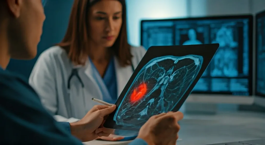
- Detailed Tumor Characterization: The MRI diagnosis of soft tissue masses can provide detailed information about a tumor’s internal composition. It can show if a growth contains fluid, fat, blood, or solid components, which helps distinguish between different types of tumors and provides clues as to whether a tumor is likely benign or malignant.
- Assessing Tumor Extent: MRI can accurately show the full extent of a tumor’s spread, including if it has invaded adjacent structures like bone or neurovascular bundles. This information is essential for surgeons planning a removal, helping them understand soft tissue tumors fully and determine a safe surgical margin.
- Differentiation from other Conditions: Soft tissue tumors can mimic other conditions, such as hematomas or infections. MRI for cancer detection provides a way to distinguish between these possibilities by revealing specific characteristics like a capsule, internal structure, and flow dynamics, leading to a more accurate diagnosis.
Benefits of MRI for Patients
Receiving an MRI for soft tissue tumors provides patients with several significant benefits, making it the preferred imaging modality for this type of condition. The procedure is painless and offers high-quality diagnostic information without the risks associated with certain other imaging methods.
Non-invasive and painless
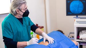
The MRI procedure is entirely non-invasive, meaning it involves no needles, incisions, or surgical intervention. During the scan, a patient lies comfortably on a table that slides into a large, tube-like machine. The process involves no physical discomfort, though some patients may feel claustrophobic inside the scanner, but this can be managed with sedation or open MRI machines.
No radiation exposure
A major advantage of MRI over other imaging techniques like CT scans or X-rays is that it does not use ionizing radiation. This makes it a very safe option, especially for patients who may require multiple scans over time for monitoring purposes. The absence of radiation exposure is a key reason many doctors recommend MRI vs CT for soft tissue tumors.
High accuracy in soft tissue visualization
MRI provides exceptionally detailed images of soft tissues, allowing radiologists to accurately characterize masses. The ability to visualize the fine details of a tumor’s structure and its relationship to surrounding tissues contributes to a very high diagnostic accuracy. This level of detail helps prevent misdiagnosis and ensures the most appropriate treatment plan is developed.
Helps guide treatment planning and surgery decisions
The detailed information from an MRI is invaluable for a surgical or oncology team. It helps them determine the best approach for a biopsy or surgical removal, identifying key structures to avoid and planning the safest, most effective way to remove the mass. It also helps clinicians determine if a tumor is resectable or requires other forms of treatment.
What to Expect During an MRI for Soft Tissue Tumors
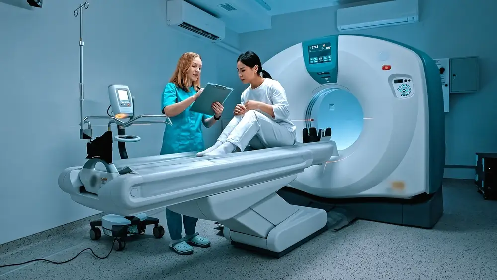
An MRI scan for lumps is a simple and straightforward procedure that takes about 30 to 60 minutes. Before the scan, you may be asked to remove any metal objects from your body, such as jewelry, watches, or piercings, as these can interfere with the magnetic field.
- The Procedure: You will lie on a padded table that slides into the MRI machine. The technologist will provide you with earplugs or headphones to help muffle the loud knocking sounds produced by the machine during the scan.
It is essential to remain as still as possible during the entire procedure to ensure the images are clear and sharp. You may be given a contrast agent through an IV to make specific tissues or blood vessels more visible.
- What You Will Feel: The procedure is painless. You will hear a series of loud, rhythmic noises as the machine works.
The most important thing is to relax and stay still. If you have any concerns about claustrophobia, inform the technologist beforehand so they can discuss options like an open MRI or light sedation.
The Clear Path Forward: High-Quality MRI for Diagnosis
An accurate and detailed diagnosis is the most important step in the treatment of soft tissue tumors. From understanding whether a growth is benign or malignant to pinpointing its exact location and size, the information gained from an MRI is a critical guide for clinicians and patients alike.
This diagnostic tool provides a safe and highly effective way to gain a comprehensive understanding of a soft tissue mass.
For those seeking a trusted and high-quality MRI diagnosis of soft tissue masses, it is important to find a facility with experienced technicians and advanced imaging technology. A reliable diagnosis begins with a clear image, which is why choosing a top-tier provider is so important.
One Step Diagnostic is dedicated to offering trusted and quality MRI services that provide the clarity needed for a precise diagnosis and effective treatment planning. Contact us today to learn more about our advanced MRI services and how we can help you take the first step towards a clear path to health and well-being.